Maria Znamenska
Associate Professor of Ophthalmology, Retina Imaging Expert
Chief Medical Officer at Altris AI
Reading time
20 min read
The burden of timely diagnostics lies on the shoulders of eye care specialists: ophthalmologists and optometrists worldwide. According to the International Agency for the Prevention of Blindness, over 1 billion people live with preventable blindness because they can’t access the proper diagnostics and treatment. Almost everyone needs access to eye care services during their lifetime. Unfortunately, there are only 331K optometrists worldwide, while 14M optometrists are required to provide effective and adequate eye care services.
With the high prevalence of the population that needs eye care services and the lack of specialists, the goal of timely and accurate diagnostics and treatment seems unachievable.

See how AI for OCT works
However, the empowerment of eye care specialists with Artificial Intelligence (AI) can be a real solution to this problem. As the larger part of the work of eye care specialists relies on retina image assessment and analysis, the support of this process can unburden ophthalmologists and optometrists all over the world. Modern AI image interpretation algorithms, such as Altris AI, can discover patterns among millions of pixels with high speed, accuracy, and zero human errors because of tiredness.
You can watch a short video of how Altris AI can assist you in detecting pathological signs on the OCT scans:
https://www.youtube.com/watch?v=Ehhwl6Q0O-A&ab_channel=Altris
In this article, we will talk about the capabilities of AI image interpretation for Optical Coherence Tomography (OCT) in detecting common pathologies, such as AMD or glaucoma, and less prevalent, such as Choroidal Melanoma. Despite the skepticism of the eye care community towards AI, multiple research works mentioned in this article prove the efficiency of AI. Moreover, there are market tools, capable of detecting 49 eye pathologies with 91% accumulative accuracy. Altris, the SaaS created by a team of retina experts based on 5 million OCT scans obtained in 11 clinics, is such a tool.
AI image interpretation for OCT
AI image interpretation for Asteroid Hyalosis
Asteroid hyalosis is a clinical condition in which calcium-lipid complexes are suspended throughout the vitreous collagen fibrils. Although it is a rare disease (1.2% prevalence according to the U.S. Beaver Dam Eye Study), it also may lead to unpleasant consequences, such as surface calcifications of intraocular lenses. Today OCT can help with the detection of this degenerative condition. For a higher confidence level, eye care specialists may use Altris AI image interpretation for OCT analysis to detect asteroid hyalosis.
AI for Central Retinal Artery Occlusion (CRAO)
Central Retinal Artery Occlusion (CRAO) presents as unilateral, acute, persistent, painless vision loss. It can be bilateral in 2% of the population. The vision loss is abrupt, and the treatment is only effective during the first hours. CRAO resembles a cerebral stroke. Therefore, its treatment should be similar to any acute event treatment: detecting the occlusion site and ensuring it won’t occur again. AI image interpretation models, such as Altris AI, can assist eye care specialists in detecting CRAO today.
AI for Central Retinal Vein Occlusion (RVO)
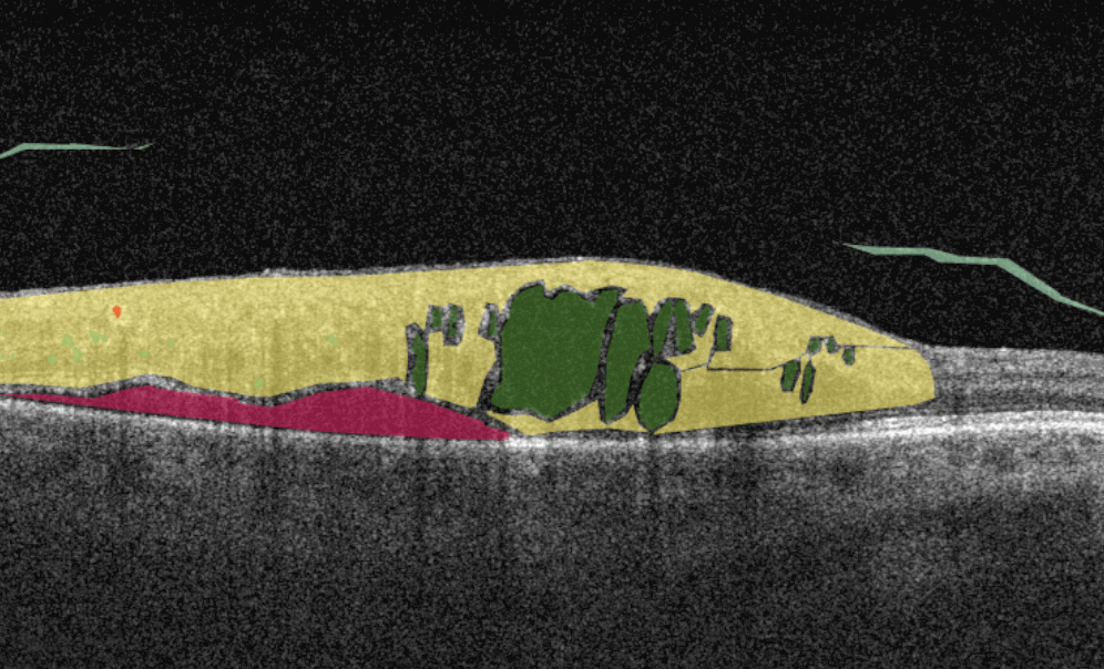
CRVO is one of the most widespread vascular diseases that affect the population over 45. There are two distinct types of CRVO: perfused (nonischemic) and nonperfused (ischemic). Each of these types has its symptoms and treatment prognosis. For instance, ischemic CRVO leads to sudden visual impairment, while nonischemic CRVO development takes time to develop mildly. The detection of CRVO is now done with the help of OCT predominately, and AI image interpretation systems shows promising results in spotting its symptoms, such as nonperfusion. Altris AI system defines CRVO with 91+% accumulative accuracy in detecting pathological signs that indicate the CRVO.
AI for Central Serous Chorioretinopathy (CSC)
Accumulation of fluid under the central retina is called central serous chorioretinopathy. Over time, this disease can lead to the distortion of vision. Fortunately, available AI models for OCT scan analysis show high accuracy in detecting CSC. This and other AI image interpretation models effectively discriminate between acute and chronic CSC, and their performance can be comparable to the performance of ophthalmologists. Altris AI is already helping eye care specialists worldwide to diagnose CSC cases.

AI for Chorioretinal Scar
Chorioretinal scars are tiny scars in the back of the eye, the size of which may vary from 0,5mm to 2mm. In most cases, the chorioretinal scar appears as the result of virus infection, such as toxoplasmosis and toxocariasis, or trauma. It usually has no malignant potential. Modern AI image interpretation algorithms allow ophthalmologists and optometrists to diagnose chorioretinal scars more accurately by relying on OCT images.
AI image interpretation for Chorioretinitis
The inflammation of the choroid is called chorioretinitis. Often, the inflammatory process can be caused by congenital viral, bacterial, or protozoan infections. Chorioretinitis is characterized by vitreous haze, fine punctate gray to yellow exudation areas, pigment accumulation along the optic nerve and blood vessels, and flame-shaped hemorrhages with chorioretinal edema. The goal of the eye care specialist is to detect chorioretinitis which can potentially lead to blindness, and to eliminate inflammation. Altris AI image interpretation system can be an excellent decision-making support tool in detecting chorioretinitis.
AI image interpretation for Choroidal Melanoma
Today, choroidal melanoma is the second most common intraocular tumor in the adult population. Patients with choroidal melanoma don’t have distinct symptoms but can have impaired visual acuity, visual field defects (scotomas), metamorphopsia, photopsia, and floaters.
OCT is a relatively new method for the detection of choroidal melanoma, which is nevertheless gaining popularity. OCT cannot be the only diagnostic method for melanoma detection – FA is also needed for final diagnosis. However, optical shadowing, thinning of overlying choriocapillaris, subretinal fluid, retina local elevation, subretinal lipofuscin deposits, and disrupted photoreceptors can be detected with the help of OCT.
Such pathological signs will indicate possible choroidal melanoma. Altris AI image interpretation system can assist eye care specialists with detecting pathological b-scans and locating this disease.
AI for Choroidal Neovascularization (CNV)
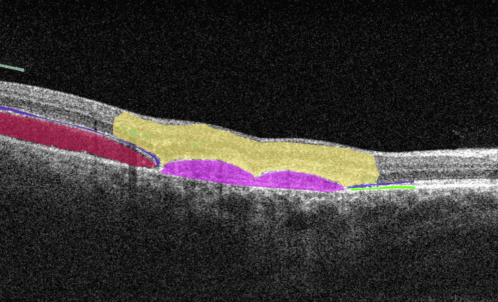
Choroidal neovascularization (CNV) is part of the spectrum of exudative age-related macular degeneration (AMD) and some other conditions. CNV is an abnormal growth of vessels from the choroidal vasculature to the neurosensory retina through Bruch’s membrane.
Modern OCT systems can detect even a tiny amount of fluid leaking into the retina. Empowered by Altris AI image interpretation algorithm, eye care specialists can spot pathological signs of choroidal neovascularization much faster or detect the pathologies that accompany CNV, resulting in better patient outcomes.
AI image interpretation for Choroidal Rupture
Traumatic choroidal rupture is common after blunt ocular trauma (5 to 10%). It is a defect in the Bruch membrane, the choroid, and the retinal pigment epithelium. The location of the choroidal rupture will define the symptoms: if the fovea and parafoveal retina are included in the rupture area, patients experience impaired vision. In other cases, the rupture can be asymptomatic. OCT is used to diagnose choroidal rupture as it can show the loss of continuity of the RPE layer and the thinning of the choroid. AI image interpretation is exceptionally accurate in layers segmentation and volume/area calculation, so missing the symptom of choroidal rupture with AI is almost impossible.
AI image interpretation for Choroidal Nevus
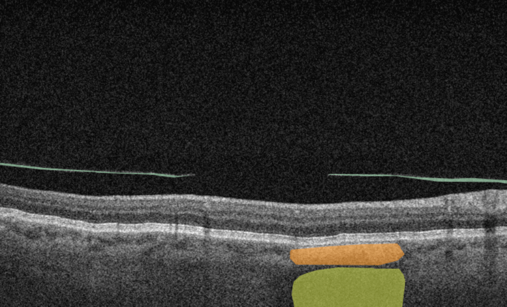
Choroidal nevus is a benign melanocytic tumor of the choroid and is found in 5 to 30% of white people. It can be found accidentally because it is asymptomatic. Artificial intelligence methods are used not only for identifying choroidal nevus but also for early signs of its transformation into malignant melanoma. The earlier the small melanoma is detected, the better the treatment prognosis is for the patient. Altris AI image interpretation system is one of the systems capable of detecting choroidal nevus before its transformation into melanoma.
AI for Cone-Rod Dystrophy (CORD)
CORD is an inherited retinal disease caused by a genetic mutation characterized by cone photoreceptor degeneration. It may be followed by subsequent rod photoreceptor loss. CORD symptoms include loss of central vision, photophobia, and progressive loss of colored vision. OCT diagnostics help to diagnose CORD by pointing at the absent interdigitation zone and progressive disruption and loss of the ellipsoid zone (EZ). Today AI image interpretation is helping to detect Cone-Rod Dystropthy to eye care specialists more confidently, even in controversial cases.
AI for Cystoid Macular Edema (СME)
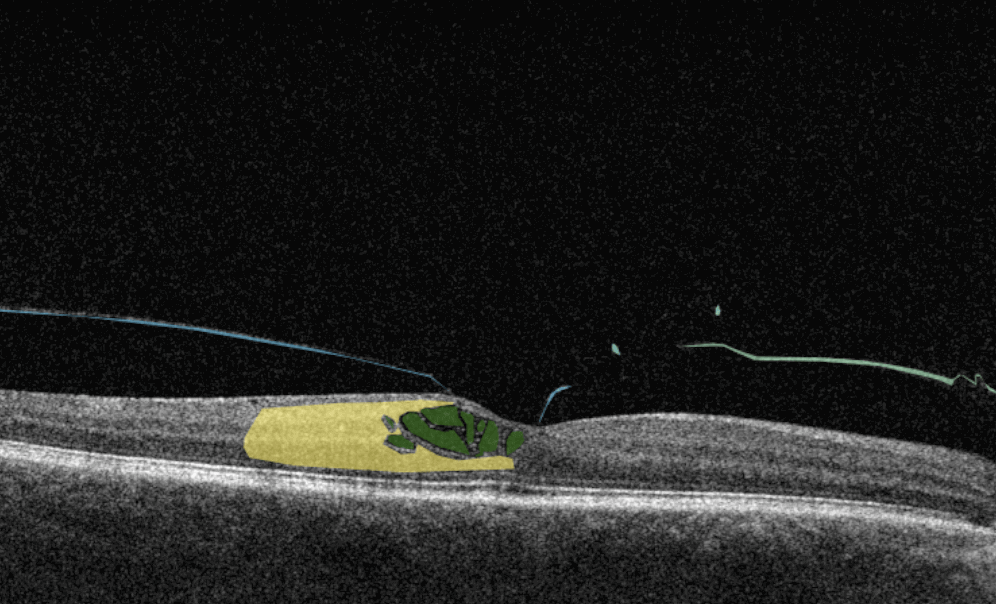
Cystoid macular edema (CME) is a painless condition in which cystic swelling or thickening occurs of the central retina (macula) and is usually associated with blurred or distorted vision. CME can be caused by many factors, including diabetic retinopathy and age-related macular degeneration (AMD). OCT diagnostics help to spot СME by detecting retinal thickening with the depiction of the intraretinal cystic areas. CME is not irreversible. Vision loss caused by macular edema can be reversed if detected early. Combining OCT diagnostics with AI image interpretation, eye care specialists can detect CME with higher accuracy at an earlier stage.
AI image interpretation for Degenerative Myopia
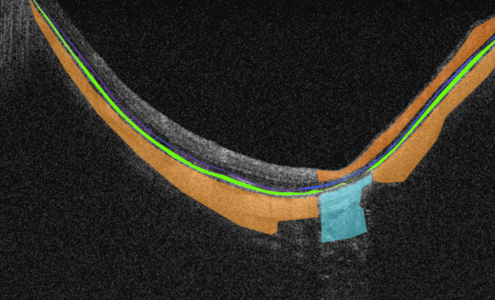
Degenerative or pathological myopia is the condition during which axial lengthening occurs, especially in the posterior pole. It leads to retina stretching, the sclera’s thinning, choroidal degeneration, and potential loss of vision. AI image interpretation systems have demonstrated excellent results in detecting pathologic myopia and identifying myopia-associated complications on OCT. AI helps ophthalmologists improve the monitoring of pathology treatment and classify different cases of myopia.
AI for Diabetic Macular Edema
Diabetic macular edema (DME) is the presence of excess fluid in the extracellular space within the retina in the macular area, typically in the inner nuclear, outer plexiform, Henle’s fiber layer, and subretinal space. DME can develop during any stage of diabetic retinopathy in patients with diabetes.
Unfortunately, the early symptoms of DME can be unnoticeable or include impaired vision and reading and color perception problems, which some people may ignore. Taking into account its asymptomatic nature, patients with diabetes need regular OCT examinations to determine the presence of DME. OCT has become a golden standard in DME detection within the last few years, and AI can be an excellent decision-making support tool in OCT scans interpretation. According to recent research, AI-powered OCT analysis provides an accurate diagnosis of DME with a cumulative accuracy of over 92%.
AI image interpretation systems can unburden ophthalmologists and optometrists who have a lot of patients due to their convenience and can be used in remote regions of the world in the future.
AI image interpretation for Diabetic Retinopathy
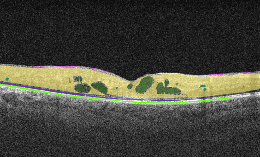
Diabetes can affect the eyes in various ways, most commonly corneal abnormalities, glaucoma, iris neovascularization, cataracts, and neuropathies. However, diabetic retinopathy (DR) is the most common and potentially the most blinding of these complications. Early treatment of both proliferative and non-proliferative DR can improve patient outcomes significantly. OCT is a common diagnostic method for diabetic retinopathy. It relies on the localization of intraretinal and/or subretinal fluid and can help to diagnose diabetic retinopathy through pathological signs detection and layer thickness measurement.
AI image interpretation is a step in the future of detection that shows high sensitivity in identifying DR, and studies prove its effectiveness. AI-assisted analysis of OCT scans helps eye care specialists today and will definitely be more widespread tomorrow.
AI image interpretation for Dry AMD
Dry AMD is a more common type of AMD (80% of people have this type), during which patients slowly lose their central vision. It is the aging of the macula and the appearance of deposits called drusen. There is no treatment for Dry AMD yet. However, early detection can help patients to change their lifestyles and slow down the development of this disease. Modern AI solutions make it possible to diagnose Dry AMD faster and develop successful methods of treating the disease. AI image interpretation systems also exclude the possibility of human error.
AI for Dry AMD – Geographic Atrophy
Geographic atrophy is an advanced form of the late stage of Dry AMD development. In this condition, retina cells will degenerate and finally die, leading to the patient’s central vision loss.
AI image interpretation is being widely used to detect Geographic Atrophy with the help of OCT. In this meta research, there are numerous studies that focus on lesion segmentation, detection, and classification of geographic atrophy and even its prediction. They vary in accuracy, but the overall trend of AI for geographic atrophy detection is very positive. The use of artificial intelligence has several advantages, including improved diagnostic accuracy and higher processing speed.
AI for ERM or Epiretinal Fibrosis

Epiretinal fibrosis (epiretinal membrane or macular puckering) is a treatable cause of visual impairment. It is a macula disease caused by fibrous tissue growth on the retina surface. AI image interpretation model for detecting ERM on OCT can outperform non-retinal eye care specialists with a cumulative accuracy of 98+%. For more professional retina experts, AI can be a decision-support tool. Early detection and treatment of this disease are crucial to prevent the growth of fibrous tissue and the worsening of the patient’s condition.
AI image interpretation for Epiretinal Hemorrhage
Epiretinal hemorrhages result from a serious trauma: car or sports accidents, falls, and direct physical impact. Mild hemorrhages unrelated to a serious traumatic event can disappear on their own, but they can be a symptom of a more complex pathology. Epiretinal hemorrhages can be detected with the help of OCT, and AI image interpretation systems can make this process more accurate.
AI for MTM (Foveoschisis)
Myopic foveoschisis or myopic traction maculopathy is the thickening of the retina that reminds schisis in patients with high myopia with posterior staphyloma.
Untreated foveoschisis often leads to vision loss due to secondary complications, which is why this disease should be detected in time. Today when OCT is becoming more widespread, detection of foveoschisis is more common and accurate. More than that, the studies show that combining the power of AI image interpretation and OCT diagnostics for MTM detection is equal to the junior ophthalmologist’s knowledge. Using AI-powered OCT, it is possible to deal with the shortage of specialists that can guarantee timely diagnostics.
AI for Full-thickness Macular Hole
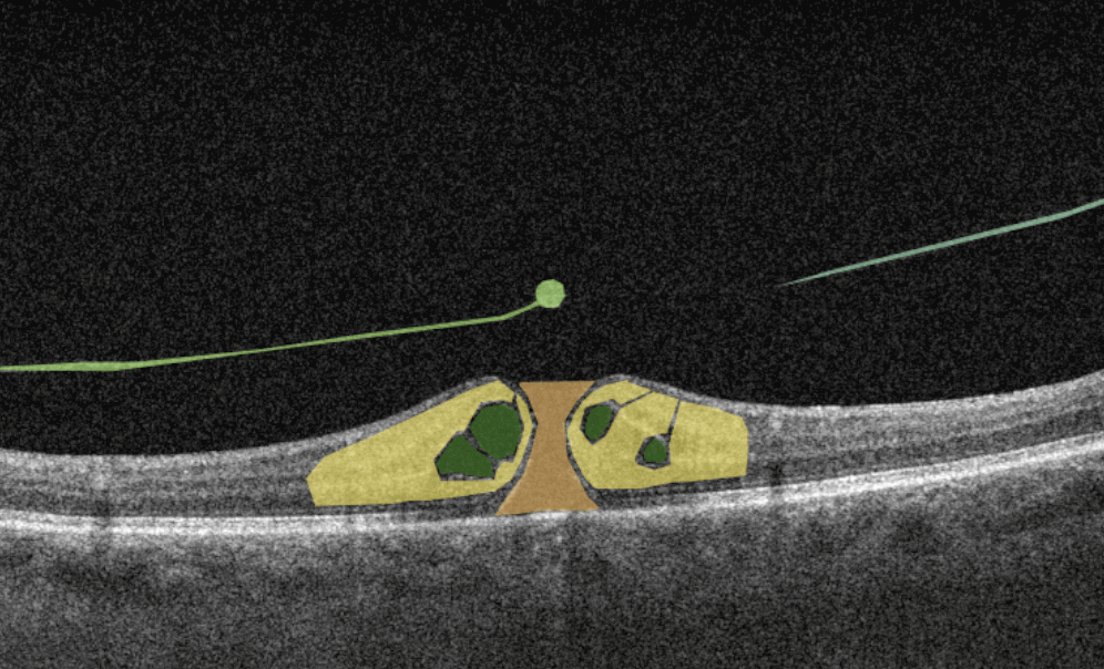
A macular hole is a full-thickness defect of the retina involving the foveal region. Patients usually present a reduction of central visual acuity. A complete ophthalmic examination, including OCT, should be performed to diagnose a full-thickness macular hole. So far, the research of AI image interpretation algorithms for a full-thickness macular hole is dedicated to OCT(A), but there are available tools on the market that can help define full-thickness macular hole on OCT scans as well. Altris AI is one of them.
AI for Hypertensive Retinopathy
People with high blood pressure, older people, and patients with diabetes often develop hypertensive retinopathy. OCT examination can be used for the detection of hypertensive retinopathy.
AI-based OCT analysis shows promising results in detecting hypertensive retinopathy by defining retinal vessels and other pathological signs in the retina.
AI for Intraretinal Hemorrhage
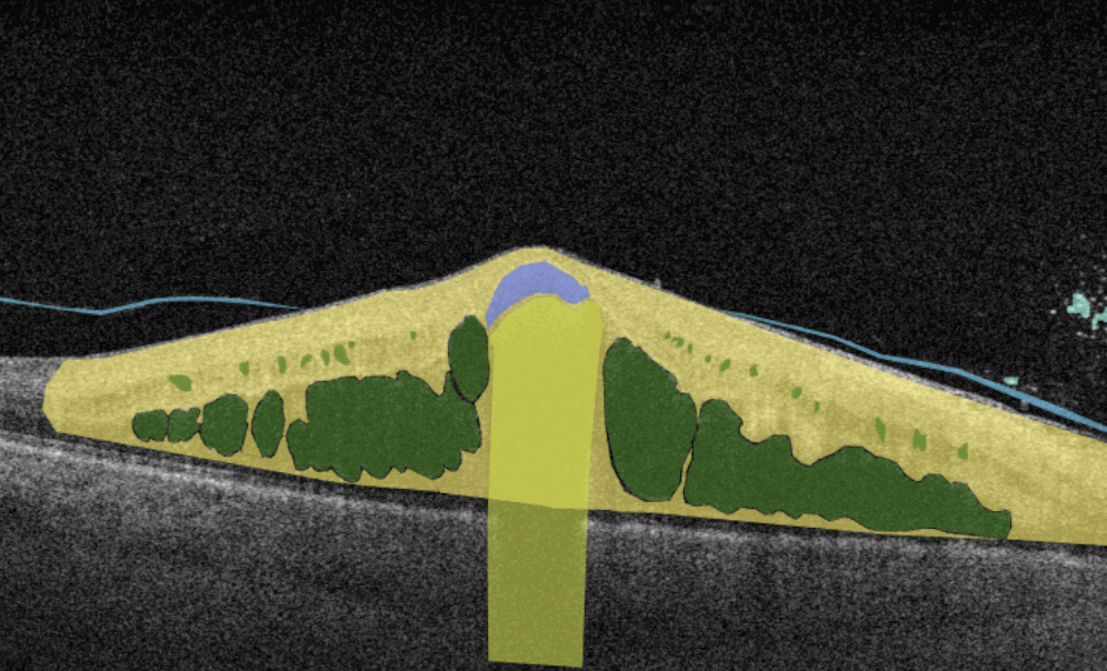
Among patients with DR, RVO, or ocular ischemic syndrome, there are often those who develop side pathologies. One of these pathologies is intraretinal hemorrhage. AI image interpretation systems help ophthalmologists and optometrists identify intraretinal hemorrhages in the retina.
AI image interpretation for Vitreous Hemorrhage
Vitreous hemorrhage results from bleeding into one of the several potential spaces formed around and within the vitreous body. This condition can follow injuries to the retina and uveal tract and their associated vascular structures. Eye care specialists should perform a complete eye examination, including OCT, slit lamp examination, intraocular pressure measurement, and dilated fundus evaluation. Timely diagnosis and treatment are essential: it can significantly reduce concomitant diseases of intravitreal hemorrhage. AI image interpretation systems can help eye care specialists detect vitreous hemorrhage supporting them in case of controversial OCT scans.
AI for Lamellar Macular Hole (LMH)
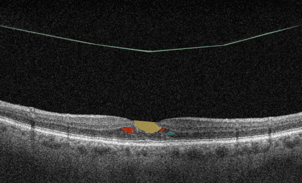
Lamellar macular hole is one of the types of macular holes known in eye care practice. The problem is that the stage 0 macular hole is a clinically silent finding detected on OCT where a parafoveal posterior hyaloid separation is present and a minimally reflective preretinal band is obliquely inserted at one end of the fovea. Eye care specialists may have problems identifying lamellar macular hole on OCT. That is where AI image interpretation models can come into play.
AI for Laser-induced Maculopathy
Since 2014, the number of laser injuries reported worldwide has more than doubled because of the widespread use of laser technologies. Depending on the damage, the patient may have a quick recovery or long-term vision loss with the development of diseases such as photoreceptor’s damage, macular hole, ERM, or others. OCT is one of the methods that help to detect laser-induced maculopathy without human errors and doubts. AI image interpretation models have a reasonable prospect of helping eye care specialists define laser-induced maculopathy based on OCT scans.
AI for Age-related Macular Degeneration (ARMD)
Age-related macular degeneration is one of the leading reasons for blindness in people of older age, especially among women and people with obesity. Patients usually present with a gradual, painless vision loss associated with delayed dark adaptation, severe metamorphopsia, and field loss. In other words, in the early stages of AMD, patients may not have any signs or symptoms, so they may not even know they have the disease. Regular OCT screening (among other diagnostic methods) can be a life-saving vest for older people.
AI image interpretation has shown great promise in detecting AMD, and research papers show that its capabilities are similar to those of ophthalmologists. AI-powered automated tools provide significant benefits for AMD screening and diagnosis.
AI for Macular Telangiectasia Type 2
Macular telangiectasia (Mac Tel) results from the capillaries abnormalities of the fovea or perifoveal region related to the retina nuclear layers and ellipsoid zone.
Macular Telangiectasia Type 2 can have negative consequences and develop into cystic cavitation-like changes in all the layers of the retina or even transform into a full-thickness macular hole. OCT is an effective diagnostic method of macular telangiectasia type 2 as the tomograph can localize foveal pit enlargement. Which is a result of secondary loss of the outer nuclear layer and ellipsoid zone that can progress into large cysts (often called ‘cavitation’) that can encompass all retinal layers.
Automating the detection of macular telangiectasia type 2 with the help of AI image interpretation systems for OCT scan analysis is already possible thanks to Altris AI.
AI for Myelinated Retinal Nerve Fiber Layer
Myelinated nerve fiber layer (MRNF) is a disease that occurs in 1%. It is a benign clinical condition that results from an embryologic developmental anomaly whereby focal areas of the retinal nerve fiber layer fail to lose their myelin sheath.
OCT is an effective method of MRNF detection with the help of the detection of the RNFL layer. Such tools as Altris AI image interpretation models are even more accurate in retina layers detection and volume measurement thanks to their growing level of accuracy.
AI image interpretation for Myopia
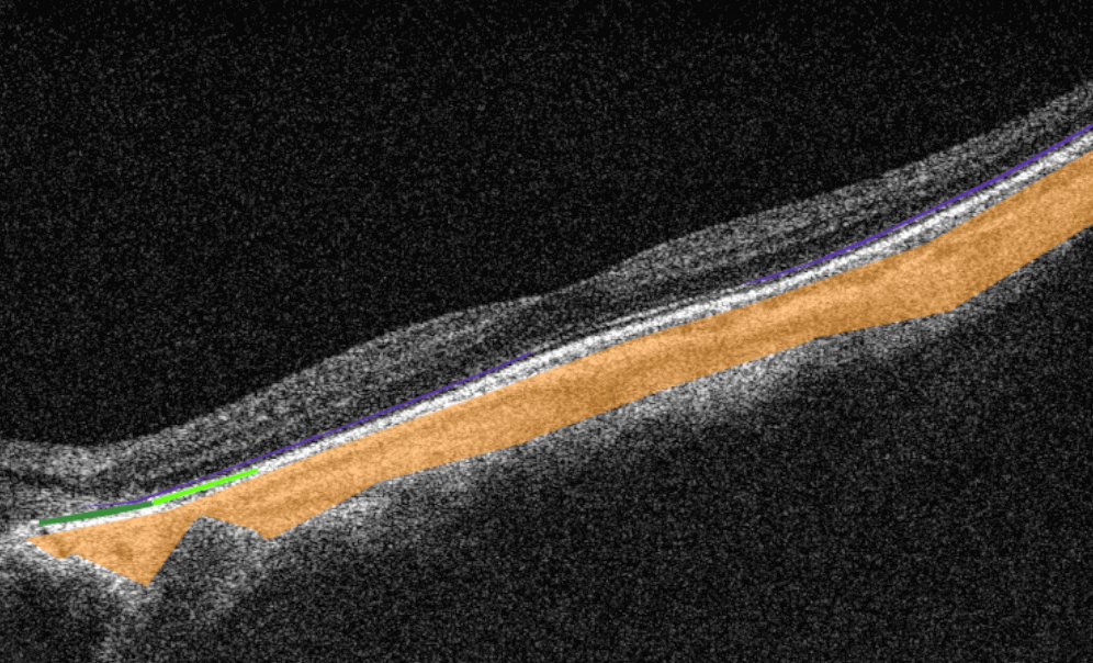
Myopia is not an eye disease. It is an eye-focusing disorder that affects 25% of the world population at a younger age. There are 2 distinct types of myopia: pathological and non-pathological — each of the types has its symptoms and treatment prognosis. The visual function of the patients, as well as the high quality of life, can be preserved if myopia is detected early enough and treated appropriately. Myopia is often diagnosed by ophthalmologists and optometrists with the help of OCT, thanks to its fine cross-sectional imagery of retinal structures. Unlike biomicroscopy, angiography, or ultrasonography, OCT can reveal undetectable retinal changes in asymptomatic patients with myopia.
Current AI image interpretation models show great promise in detecting myopia on OCT scans, and their results can be compared to the results of junior retina specialists. Altris AI is an accurate AI tool for myopia.
AI for Pigment Epithelium Detachment
Retinal pigment epithelial detachment (PED) is often observed in Wet AMD and other conditions. It is determined as a separation of the RPE layer from the inner collagenous layer of Bruch’s membrane. With its capability to visualize retinal layers, OCT helps eye care specialists with timely PED diagnostics. Powered with AI image interpretation systems, OCT diagnostics can promise zero human errors and exceptional accuracy.
AI for Polypoidal Choroidal Vasculopathy (PCV)
Polypoid Choroidal Vasculopathy is a disease of the choroidal vasculature. Serosanguineous detachments of the pigmented epithelium and exudative changes that can commonly lead to subretinal fibrosis are the main OCT signs of PCV. AI image interpretation systems show great potential in establishing a difference in diagnostics between PCV and AMD.
AI for Preretinal Hemorrhage
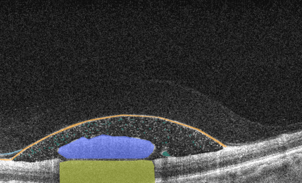
Preretinal hemorrhage is a complication of many pathologies, such as leukemia or ocular/head trauma. Missing preretinal hemorrhage means putting a patient at risk. Preretinal hemorrhage can be a presenting sign of some systemic diseases. In any case, OCT diagnostics are performed to determine preretinal hemorrhage and its real reason.
AI image interpretation for Pseudohole
Sometimes the pulling or wrinkling of the epiretinal membrane (ERM) can result in a gap called a pseudohole. A pseudohole can look like a macular hole; sometimes, it can turn into one, so it is essential to distinguish between these two phenomena. Optical coherence tomography can accurately determine a pseudohole revealing an epiretinal membrane with contraction of the retina or suppression of retinal layers. Combined with AI image interpretation, OCT diagnosis can guarantee higher accuracy in pseudohole detection.
AI for Retinal Angiomatous Proliferation (RAP)
RAP is a subtype of AMD, which is neovascularization that starts at the retina and progresses posteriorly into subretinal space. There are 3 stages of RAP: intraretinal neovascularization (IRN), subretinal neovascularization (SRN), and choroidal neovascularization (CNV). OCT is effective for detecting IRN only since changes beneath the pigment epithelium are challenging to assess. AI image interpretation models effectively differentiate between RAP and polypoidal choroidal vasculopathy (PCV), comparable to the performance of eight ophthalmologists. AI cannot substitute eye care specialists but can be an excellent decision-making support tool.
AI for Retinal Detachment
Retinal detachment is a serious eye condition that happens when the retina pulls away from the tissue around it. It can be a result of trauma or another disease. OCT has become a new standard for detecting early retinal detachment and defining the best time for surgical operation, for example. OCT powered with AI image interpretation systems can give eye care specialists the confidence they need to determine the degree of detachment and make the correct prognosis.
AI for Retinitis Pigmentosa
RP is a hereditary diffuse pigment retinal dystrophy characterized by the absence of inflammation, progressive field loss, and abnormal ERG. OCT diagnostics allows assessing morphological abnormalities in RP, providing insights into the pathology of RP and helping to make a good prognosis. AI image interpretation applied for the OCT analysis shows promising results in Inherited Retinal Diseases detection and future management.
AI image interpretation for Retinoschisis
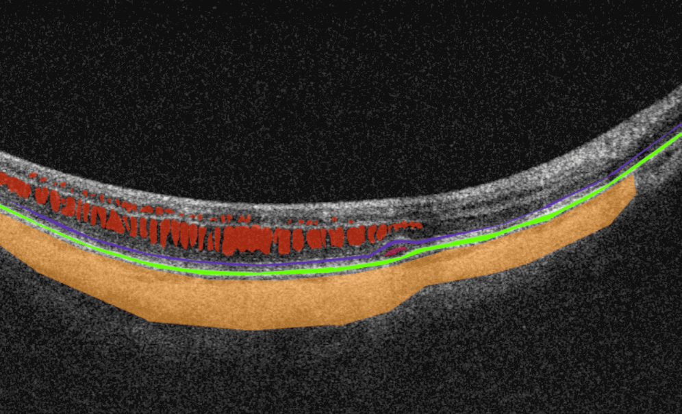
Retinoschisis is an eye condition characterized by a peripheral splitting of retinal layers. OCT is an effective method of retinoschisis diagnostic. The application of AI image interpretation tools for OCT analysis for identifying retinoschisis (among other myopia conditions) is comparable to the performance of experienced ophthalmologists.
AI for Retinal Pigment Epithelial (RPE) Tears (Rupture)
RPE rupture or RPE tears is the condition when this retinal layer acutely tears from itself and retracts in an area of a retina, usually overlying a pigment epithelial detachment (PED). OCT is an effective method of diagnostics of RPE tears. OCT scans will show a discontinuity of the hyperreflective RPE band, with a free edge of RPE usually wavy and scrolled up overlying the PED, contracted back towards the CNVM.
AI image interpretation systems can provide eye care specialists with confidence when detecting RPE tears. Systems such as Altris AI can distinguish between retinal layers with exceptional accuracy, exceeding the accuracy of eye care specialists.
AI for Solar Retinopathy (Maculopathy)
Solar retinopathy is photochemical toxicity and the consequent injury to retinal tissues located in the fovea in most cases. OCT helps to diagnose solar retinopathy by indicating changes and focal disruption at the level of the subfoveal RPE and outer retinal bands. The overall retinal architecture remains intact. AI image interpretation models can confidently assist eye care specialists in detecting solar retinopathy, even when they are in doubt.
AI for Subhyaloid Hemorrhage
Subhyaloid hemorrhage is diagnosed when the vitreous is detached from the retina because of blood accumulation. This type of hemorrhage is rare and is different from intraretinal hemorrhage caused by trauma or diabetes. OCT helps to detect subhyaloid hemorrhage. For eye care specialists who don’t have experience in detecting subhyaloid hemorrhage, the AI image interpretation model can become a great support tool.
AI for Subretinal Fibrosis
Subretinal fibrosis appears due to wound healing reaction to the choroidal neovascularization in nAMD or other conditions. Early diagnostics of subretinal fibrosis are critical because a neovascular lesion’s transformation into a fibrotic lesion can be very rapid. OCT is regarded as the most accurate method of diagnostics today.
AI image interpretation systems can help eye care specialists who use OCT with early diagnostics of subretinal fibrosis and improve patient outcomes.
AI for Subretinal Hemorrhage
Subretinal hemorrhages are a complication of various diseases which arise from the choroidal or retinal circulation. It is most often caused by AMD, trauma, and retinal arterial macroaneurysm. OCT will be an effective tool for determining the level at which subretinal hemorrhage occurred. Powered with AI image interpretation models, OCT can become the decision-making support tool eye care specialists need for subretinal hemorrhage identification.
AI for Sub-RPE (Retinal Pigment Epithelial) Hemorrhage
Sub-RPE (retinal pigment epithelium) hemorrhage is located between the RPE and Bruch’s membrane. OCT is an essential tool for validating the hemorrhage’s diagnosis and localization. AI image interpretation tools, such as Altris AI, will ensure that Sub-RPE hemorrhage is not missed.
AI for Tapetoretinal degeneration or dystrophy
Tapetoretinal dystrophy or tapetoretinal degeneration (TD) is exogenous destruction of the retina caused by a genetic mutation. Eye care specialists might easily miss such rare conditions as tapetoretinal degeneration. It is often advisable to have AI image interpretation systems as a decision-making support tool not to miss TD or other uncommon diseases.
AI image interpretation for Vitelliform Dystrophy
It is autosomal dominant degenerative maculopathy wherein a mutation in the bestrophin gene leads to lipofuscin accumulation in RPE cells manifested in a yellow spot. Detecting vitelliform dystrophy is critical at the early stages as it can lead to vision loss. OCT provides essential information on the lesion’s morphology, location, and dynamics. Empowered with AI image interpretation tools, such as Altris AI, eye care specialists won’t miss such a rare disease as vitelliform dystrophy at the early stage.
AI for Vitreomacular Traction Syndrome
Vitreomacular traction syndrome is a pathological condition characterized by a posterior vitreous detachment that leads to blurred vision or serious vision impairment. OCT is an essential method of diagnostics of vitreomacular traction syndrome as it can show the amount of involvement and tension on the macula caused by VMT. Combined with AI image interpretation tools, OCT analysis can give incredible results.
AI image interpretation for Wet AMD
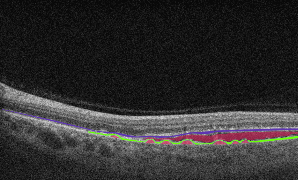
Wet AMD is the most widespread disease among the elderly population in developing countries. It is a disease characterized by abnormal blood vessel growth under the retina. Understanding that this disease can lead to rapid and severe vision loss, its early detection and treatment are very important. OCT is a golden standard for the diagnostics of wet AMD as it shows fluid or blood underneath the retina without dye, among other pathological signs.
Today AI shows promising results in predicting the development of wet AMD based on OCT images. For instance, the DARC algorithm designed for detecting apoptosing retinal cells could predict new wet-AMD activity. Another effective AI image interpretation algorithm determines the location and volumetric information of macular fluid within different tissue compartments in wet AMD, providing eye care specialists with the ability to predict visual acuity changes.
AI for X-linked Juvenile Retinoschisis (XLRS)
XLRS is a rare congenital retina disease caused by mutations in the RS1 gene, which encodes retinoschisin, a protein involved in intercellular adhesion and likely retinal cellular organization. The disease usually affects younger males in their teenage years who complain about blurred vision. OCT is used to detect schisis in the superficial neural retina and thinning of the retina. Despite the lack of research articles on AI in OCT diagnostics of XLRS, there are AI image interpretation tools that already cope with this task effectively.
Final Words
Artificial intelligence can identify, localize, and quantify pathological signs in almost every disease of the macula and retina. That is how AI image interpretation systems can provide decision-making support with the pathologies at their early stages or rare pathologies. AI can help to detect many pathologies that are invisible to the human eye because of their size or that are at their early stage.
The overall potential of artificial intelligence for ophthalmologists and optometrists is enormous and includes pathological scan selection and scan analysis with the probability of existing pathologies and pathological signs. One trial is worth a thousand words in the case of AI tools for ophthalmologists and optometrists.

