Recently Posted
-

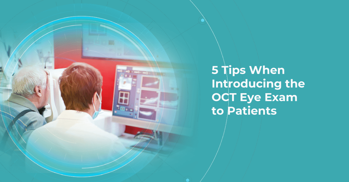
5 Tips When Introducing the OCT Eye Exam to Patients
 Mark Braddon
24.07.20238 min read
Mark Braddon
24.07.20238 min readAs optometry technology evolves, many optometrists predict that utilizing OCT eye exam in practice will be vital in maximizing patient care. That is why successfully integrating an OCT device into your optometry practice workflow is instrumental to its clinical and commercial success.
Optometrists from different countries often have the same questions about how to successfully integrate an OCT device into an Optometrist Practice, regardless of practice size or experience level. How to make patients feel comfortable? How to explain the importance of regular OCT scans? Will patients understand what is an OCT scan of the eye? How do we avoid patients thinking we want to perform OCT eye exams just to earn more money? The process of introducing OCT to patients is complex and covers many areas.
If we speak to optometry practices, both those who are new to OCT and those who have had the OCT device for many years, most of them will want to improve the ROI and ensure the patients are gaining the full value of the OCT eye test. This article will show you 5 tips for successfully introducing the OCT eye exam to your patients.
Remember why you invested in the OCT technology
One may think that only novice optometrists tend to underestimate their work or do not feel confident about the value they give to patients. However, some experienced clinicians also avoid offering OCT eye tests because they think they are ‘overselling’ with additional fees for OCT, Optos, or other diagnostic exams.
That is why it is important to remember why you invested in OCT technology in the first place. In almost all cases, this is to improve the clinical standard of eye care that you offer to your patients. In fact, when I ask some optometrists if they want a member of their families to have an OCT eye exam, the answer is always ‘Yes, of course!’. So if you strongly recommend undergoing an examination to your relatives, why would you not recommend an OCT eye test for your patients?

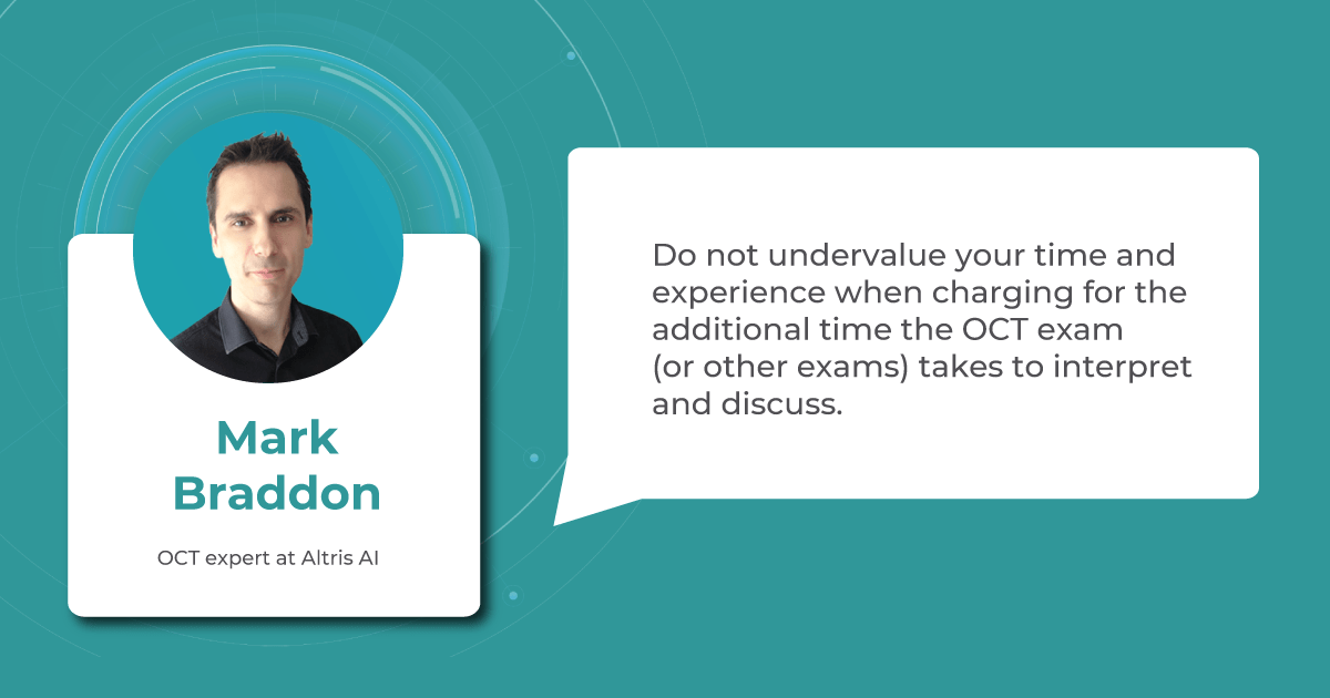
Before a patient comes into the practice, one of the most important things you need to do is not undervalue your time, skills, and experience when charging for the additional time the OCT exam takes to interpret and discuss.
Implementing an OCT eye exam into regular practice improves clinical care and can generate a commercial benefit as well by increasing revenue through fees, patient retention, and loyalty. Moreover, word of mouth is often the most significant source of new patients for optometrists. If the patient feels you are confident in everything you do, it will make them more likely to recommend you to friends and family
Explain the importance of OCT eye exam for early detection
From the first touch point, the patient should understand that your optometric practice takes its business seriously and provides additional diagnostic examinations, such as the OCT, to improve the quality of care. The first impression of your approach is very important, so it is crucial to start introducing the technology to the potential patient from the first point of contact.
As a rule, the beginning of a patient’s introduction to the OCT eye exam starts with several touch points. Whether they make their appointment for the eye examination through your website, mobile application, in person, or by phone, the most important thing you can do is create an integrated and comfortable patient journey.

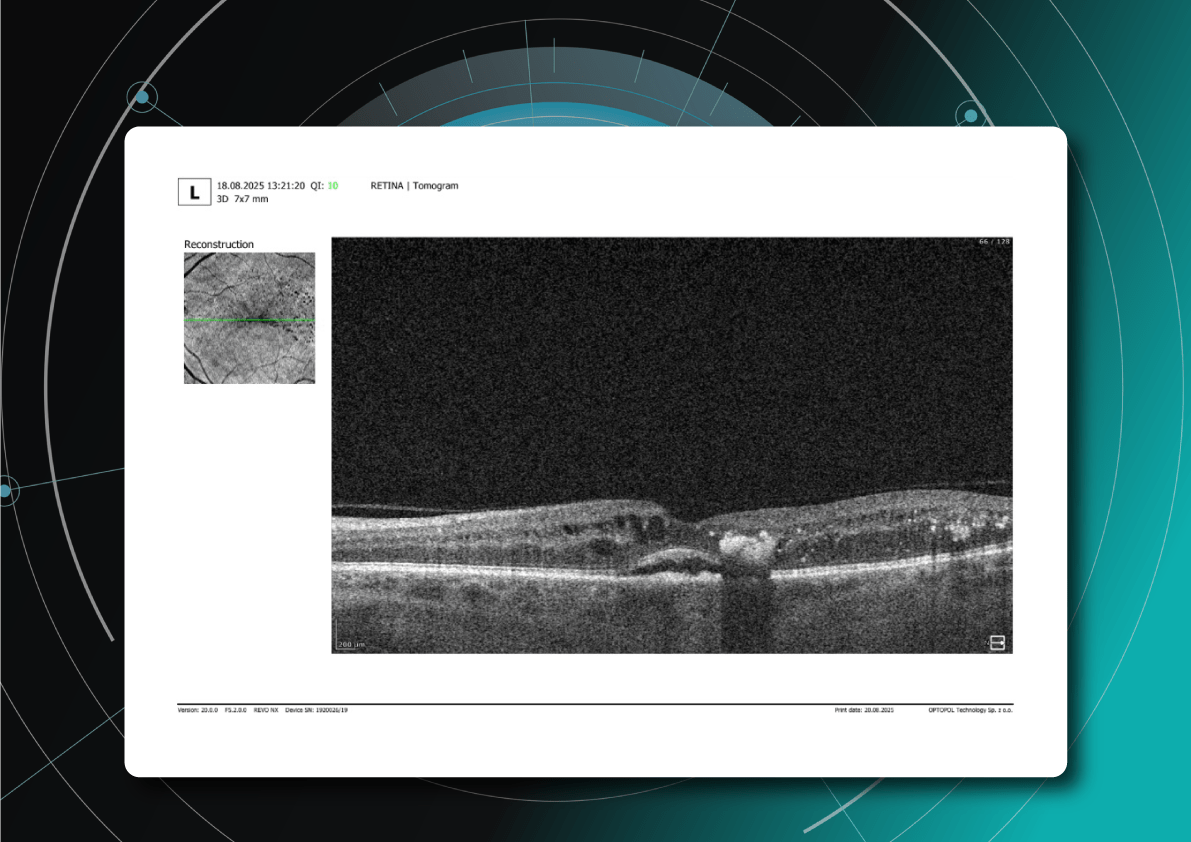
Before a patient comes into the practice, you should explain the importance of the OCT device and its benefits compared to the standard examination. Even when the patient is fully acquainted with the OCT eye exam, they may still need help understanding why this particular imaging method is necessary. The ability of OCT eye exam to detect diseases in the early stages makes this technology indispensable for optometrists and patients and this is why it is such an excellent tool for diagnosing eye diseases.
More importantly, avoid frightening patients with stories about difficult-to-treat rare pathologies. Instead of talking about the pathology consequences, say that the OCT eye exam scan provides a clear map that helps locate areas of the eye with abnormalities or early changes.
Understand the importance of a healthy-eye-as-a-baseline concept
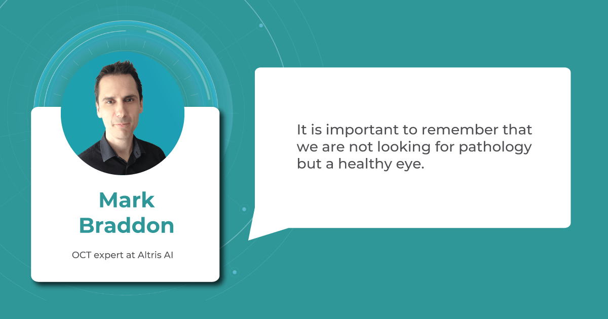
In this section, I want to discuss the concept of a healthy eye in more detail. When a patient comes to you for an examination, it is essential to use the correct narrative that the optometrist should use when discussing the results of an OCT eye exam with patients. It is important to emphasize that we are not looking for pathology but a healthy eye.
We know that we will detect pathology in certain patients. The number of patients likely to have at least one pathology increases if you work with an older population. However, finding a healthy baseline scan is an important part of monitoring the long-term eye health of the patient.

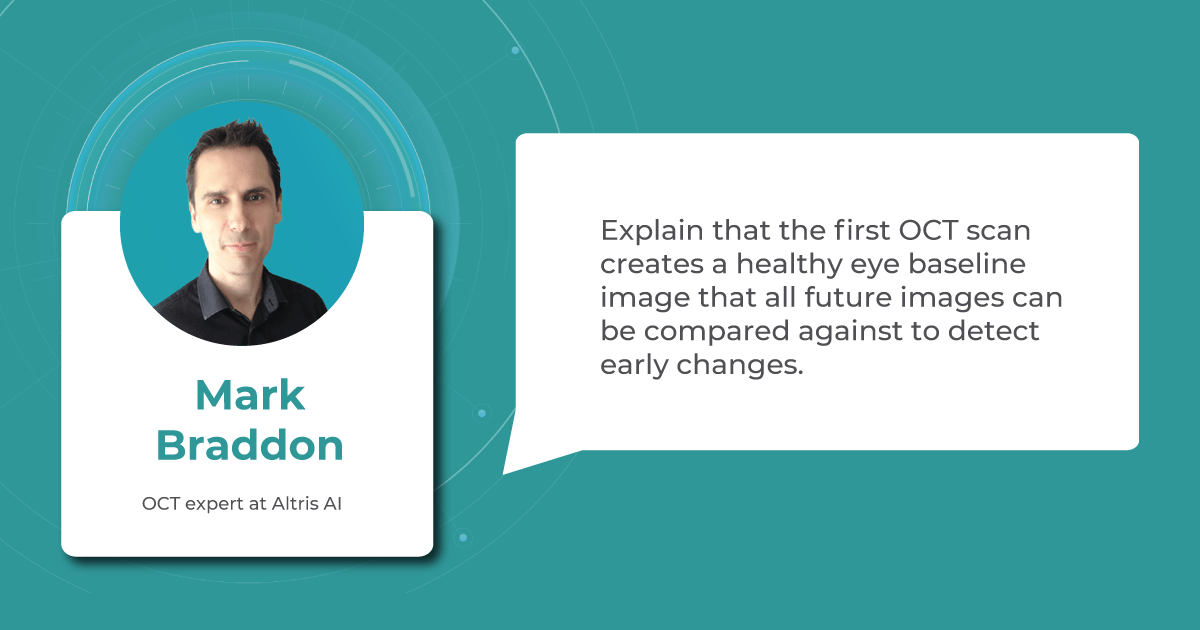
Talking about baseline, make sure to emphasize how great it is to find a healthy eye in a patient. Explain that together you found a nice, healthy eye so you have the baseline to compare with the patient’s future scans. Emphasize that, hopefully, you will find a healthy eye at the next eye examination, but if anything does start to change, then with the help of an OCT eye exam, you will be able to detect these early and minor changes as you have the healthy baseline scan to compare to.
It is necessary to develop your patient’s understanding through appropriate teaching and discussion. Giving the value of the baseline OCT eye exam to your patients is very important. Notice the difference between “We found nothing” and “We found a healthy eye”. The first statement is negative and undermines the reason for the scanning of patients for a healthy eye baseline. Meanwhile, the second statement is positive and clearly gives your patient more value as you have found what you are looking for.
Integrate the OCT eye exam into the patient workflow
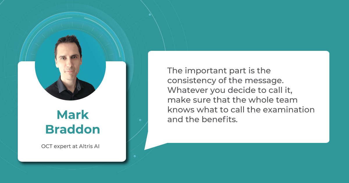
Another one of my recommendations is to call the eye examination that includes the OCT eye exam the Advanced or Comprehensive Eye Examination. It is important to make sure all the staff members use the same terminology and your message to a patient is consistent from first contact to the end of the practice visit. The eye examination without the OCT exam can be called the ‘Standard Examination’ as we are not trying to make the ‘normal’ eye examination appear below standard, what we are trying to do is explain that the practice is invested in the latest technology to offer the most advanced (or comprehensive) examination for your patients benefit.

For example, when a patient books an appointment, make sure that the support staff uses the same terminology as written throughout the website, reminder letter/email, or mobile app if you have one.
When you review the OCT images with the patient, explain that you are going to look at the OCT images of the retina, which is part of the ‘Advanced examination’. When a patient pays at the end of the customer journey, make sure that the ‘Advanced Examination’ is mentioned again. When a patient rings up or books online for the next OCT eye exam, then they will understand what the ‘Advanced examination’ means and are more likely to select this option straight away for future examinations.
Concentrate on giving more value to your patients
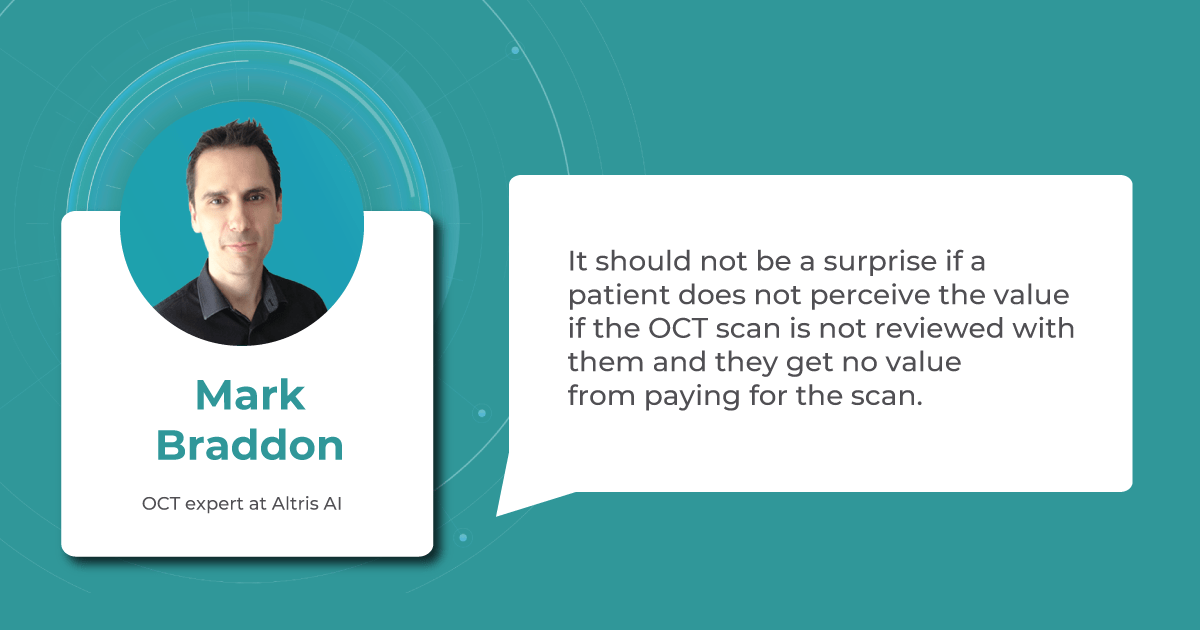
Review the results with the patient to give them the actual value of an OCT scan. This will allow you to establish communication with the patient and improve their perception. Give them the “theatre” around the additional diagnostic testing so they understand how it applies to them and feel valued.

Remember that your knowledge, enthusiasm, and the extent to which the patient is involved in the process directly affect the clinical and commercial success. Dedicate time to each patient, involve them in the diagnostic process, and explain the OCT scans of their eyes on the screen.
How can Altris AI help with introducing OCT Eye Exam

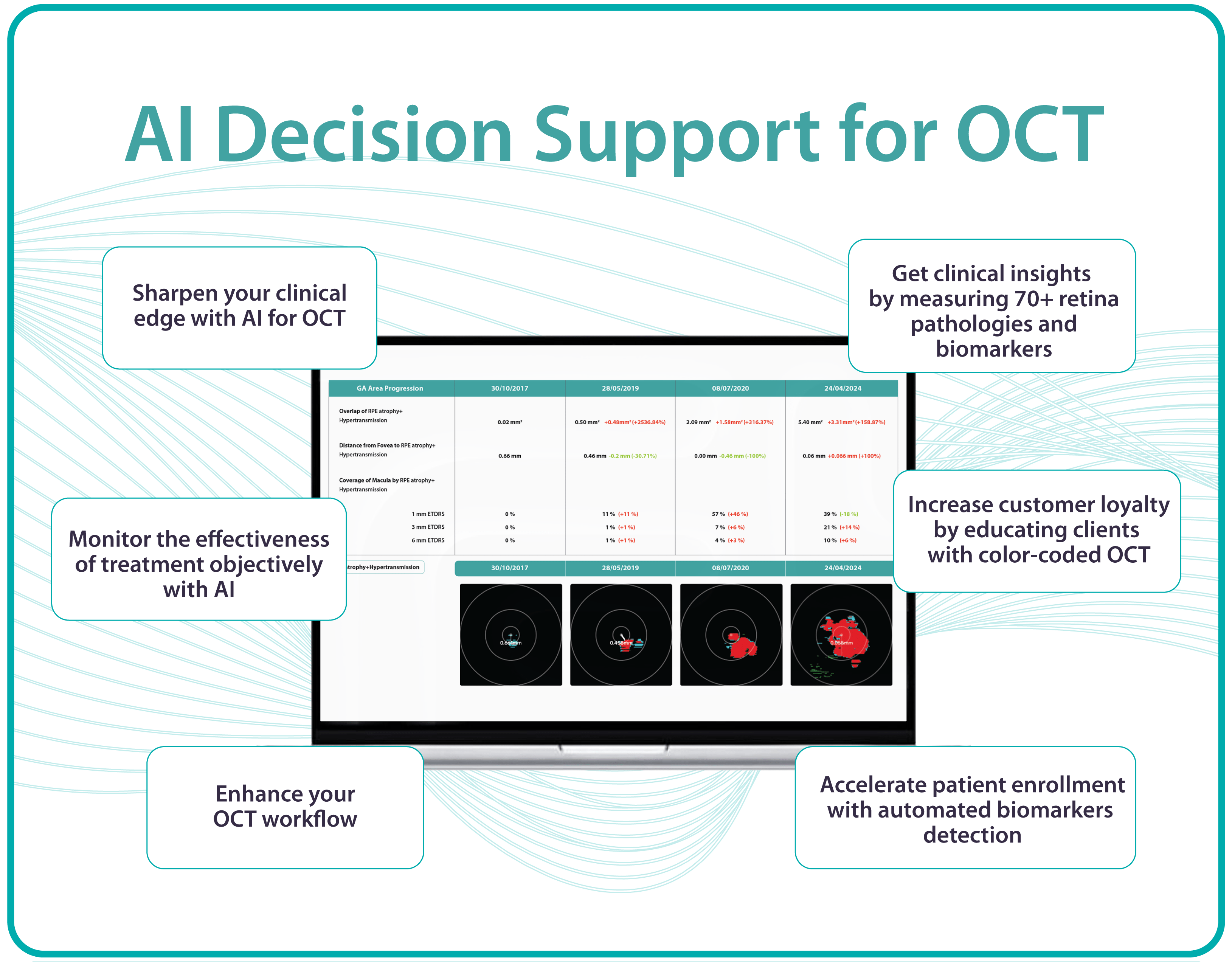
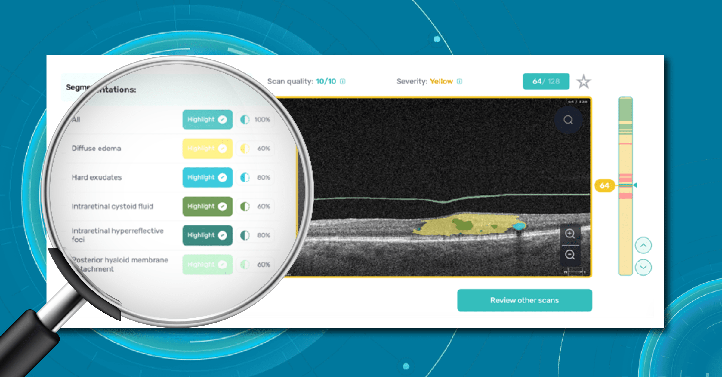
When talking about improving the clinical standard of care your practice offers to your patients, the Altris AI platform can also improve the standard of care you offer to your patients. The platform helps to quickly determine if the eye is healthy. If pathology is detected, then Altris AI identifies the very early, rare, or minor changes that can be the start of something more severe. Altris AI detects over 70 pathologies and pathological signs. If early pathology is identified, then the Altris AI platform can help educate the patient by clearly highlighting the areas of concern and then giving you the opportunity to discuss lifestyle changes, over-the-counter medications, or supplements, which may help the patient now rather than just monitoring until it is time to refer.
The Altris AI platform can improve the patient’s understanding of the OCT exam and add value to the Advanced Eye Examination.

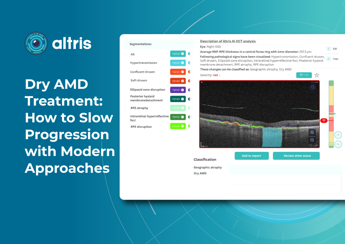
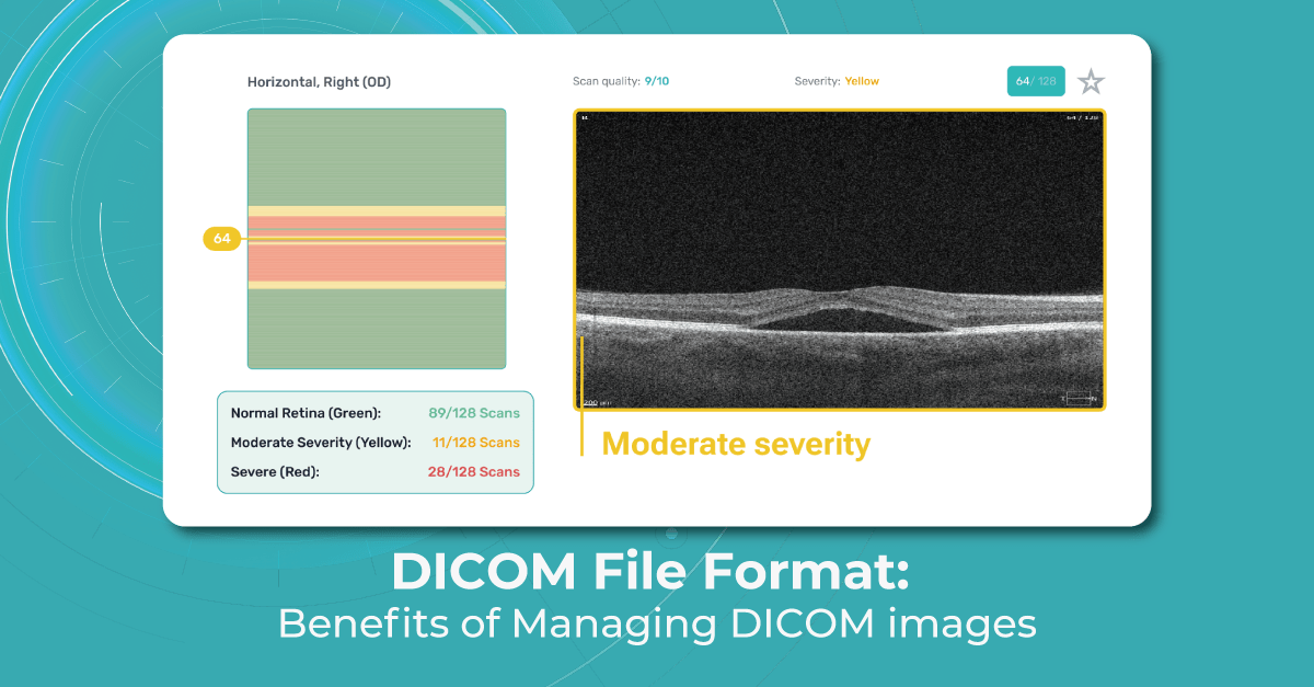
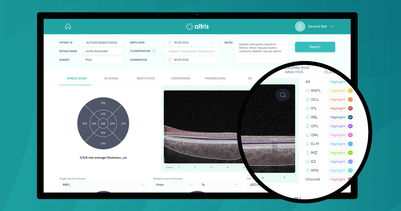
All you need to do is to upload an OCT macula exam to the platform and Altris AI will assess the exam by severity differentiating the b-scans between high, medium, and low severity levels. The segmentation/classification module will highlight pathological signs on the OCT scan one by one and give the classification/s of any pathology found to support you with the diagnosis. Meanwhile, in the Comparison module of the platform, you are able to compare the baseline scan with the current one.
Summing Up
Remember why you invested in the OCT technology in the first place — usually, this is to improve the clinical standard of care you can offer to your patients. The improvement in clinical care can also generate a commercial benefit as well by increasing revenue through OCT exam fees, patient satisfaction, patient retention and loyalty, and an increase in recommendations of friends and family.
Build a patient journey in such a way that, at each stage, they know that they have received a new, exciting, and, important part for the most comprehensive examination you offer. Remember that the more skill and enthusiasm you show, the more you can interest the patient and increase the probability that they will return for their next examination with OCT.
In addition, consider using modern AI tools to help you with decision-making. Image management systems like Altris AI can help you interpret the OCT scans faster and with more confidence. This will leave more time to add value for your patient, and integrating AI into practice can be another example of how you are investing in the latest technology to benefit your patients.
Disclaimer: USA FDA 510(k) Class II; Altris Image Management System (Altris IMS); AI/ML models and components intended to use for research purposes, not for clinical diagnosis purposes.
-

Business Case: Lux Zir and OCT Analysis in Ophthalmic Clinic
 Altris Inc.
11.07.20232 min read
Altris Inc.
11.07.20232 min readBusiness Case: Lux Zir and OCT Analysis in Ophthalmic Clinic
The Client: Lux Zir is one of the best-known ophthalmic clinics in Ukraine which provides retina diagnostics and eye treatment services. The clinic currently employs 3 full-time eye practitioners 2 general ophthalmologists and a pediatric retina expert.
The clinic normally sees between 15-20 per day with up to 10 OCT examinations performed.
The Problem:
Luxzir uses Optical Coherence Tomography as one of its core diagnostic methods because of its high level of accuracy and non-invasiveness. However, the clinic needed to solve several typical problems related to OCT.
- Some ECPs have less experience with OCT interpretation than others and this creates an inconsistent standard of care throughout the clinic.
- Some ophthalmologists come across complex OCT scans that they are unable to interpret without the help of their more experienced colleagues.
- It is difficult to maintain a high standard of care for diagnostics when the CMO is absent during the period of vacation or sick leave.
- Take out wrong and start with an inaccurate diagnosis on the basis of OCT of the patients who are referred to the clinic from other eye care centers.
The Solution:
Lux Zir Ophthalmic Clinic decided to implement the Altris AI platform as they understood how it can help resolve their problems. The results have been very positive with improvements with all issues above problems, and received very positive results.

According to Marta Shchur, Chief Medical Officer at Lux Zir clinic, the implementation of the Altris AI system improved the level of OCT diagnostics inside the clinic or if to be precise:
- OCT interpretation is now considerably faster allowing the ECPs to see 10% more patients per day.
- OCT diagnostics has become much more efficient: supported by Altris AI, ophthalmologists now have confidence when diagnosing pathologies and pathological signs, even rare ones.
- The quality of diagnostics is consistent regardless of the experience of the specialists.
Disclaimer: USA FDA 510(k) Class II; Altris Image Management System (Altris IMS); AI/ML models and components intended to use for research purposes, not for clinical diagnosis purposes.
-

Business Case: Altris for Jeff Sciberras Optometry
 Altris Inc.
10.07.20231 min read
Altris Inc.
10.07.20231 min readBusiness Case: Altris for Jeff Sciberras Optometry
The Client: Canadian Optometry Clinic
Jeff Sciberras Optometry Clinic is an established eye care facility in Mississauga, Canada. They have been recognized as the Top Choice Optometry Clinic for the past five years running in this large Canadian city.
Dr. Jeff Sciberras is proud of his high patient satisfaction rate: 92% of those surveyed would refer a friend, colleague, or family member to this establishment.
Dr. Sciberras aims to provide comprehensive eye care, with a desire to utilize leading technologies and the delivery of premium eye care products.
Recent technology investments include OCT, which allows earlier diagnosis and greater in-house management capabilities.
The Challenge: The optometry clinic has just purchased a brand new Optopol Revo OCT equipment and the support was needed in OCT scan interpretation. OCT is one of the most accurate methods of retina diagnostics however, the interpretation of OCT scans can be challenging and time-consuming, for both doctor and patient.
The Result:
Dr. Sciberras has been extremely satisfied with the support that the Altris AI platform provides:
- Increased confidence when working with the new OCT device · more profound analysis of OCT scans
- More adequate referral of complex cases.
- Scan summaries for the patient.
- Earning patient confidence and trust: The image of the innovative optometry center is enhanced to their patients and families.
- The AI Segmentation/Classification Module is invaluable for the optometry center as this module helps in the identification of 70+ pathologies and pathological signs.
The introduction of OCT with Altris AI has transformed my practice literally overnight. The integration was seamless and Altris customer support has been outstanding.
Overall, Dr. Sciberras has been impressed with the experience and support Altris AI provides and is happy to have chosen to partner with them for his leading eye care center.
- Disclaimer: USA FDA 510(k) Class II; Altris Image Management System (Altris IMS); AI/ML models and components intended to use for research purposes, not for clinical diagnosis purposes
-

DICOM Format: Benefits of Managing DICOM images
 Mark Braddon
31.05.20236 min read
Mark Braddon
31.05.20236 min readDICOM Format: Benefits of Managing DICOM images
DICOM file format (Digital Imaging and Communications in Medicine) was developed by the American College of Radiology (ACR) and the National Electrical Manufacturers Association (NEMA) as a standard for exchanging medical images and related information across different healthcare systems. It serves as a universal language for medical imaging, enabling interoperability between various imaging devices and systems. DICOM ensures that medical images can be exchanged and viewed consistently regardless of the manufacturer or modality.
DICOM image format supports a broad range of medical imaging modalities, including X-ray, MRI, OCT, ultrasound, nuclear medicine, and more. It also covers related data, such as patient information, study details, image annotations, and results
As the DICOM format continues to evolve to keep up with advancements in medical imaging technology, our article aims to raise awareness among ophthalmologists and optometrists about the DICOM file format.
What is DICOM format? You can also watch a short video about DICOM and non-DICOM file formats.
What is DICOM file format?
Image files that adhere to part 10 of the DICOM standard are commonly known as “DICOM format files” or simply “DICOM files,” and their file extension is “.dcm.” In ophthalmology, DICOM is a widely used file format for storing and transmitting medical images. DICOM files are used to store various types of ophthalmic images as well, including retinal images, optical coherence tomography (OCT) scans, visual field tests, and angiography images.
DICOM files consist of two main components: the header and the image data. The header contains metadata that describes the patient, study, series, and image acquisition parameters.

This metadata includes information such as patient demographics, image acquisition parameters (e.g., imaging modality, image orientation, pixel spacing), and any annotations or measurements made on the image. The image data itself is typically stored in a compressed format, such as JPEG or JPEG 2000, within the DICOM file.
DICOM files also support the exchange of images and associated data between different medical imaging devices and systems. This enables eye care specialists to easily share and access ophthalmic images across different platforms, such as picture archiving and communication systems (PACS), ophthalmic imaging devices, and electronic health record (EHR) systems.
By using DICOM, ophthalmologists and optometrists can efficiently store, retrieve, and analyze ophthalmic images, ensuring accurate diagnoses and effective patient care. In the next paragraphs, we will tell you more about the benefits of the DICOM file format for eye care specialists.
Benefits of DICOM format
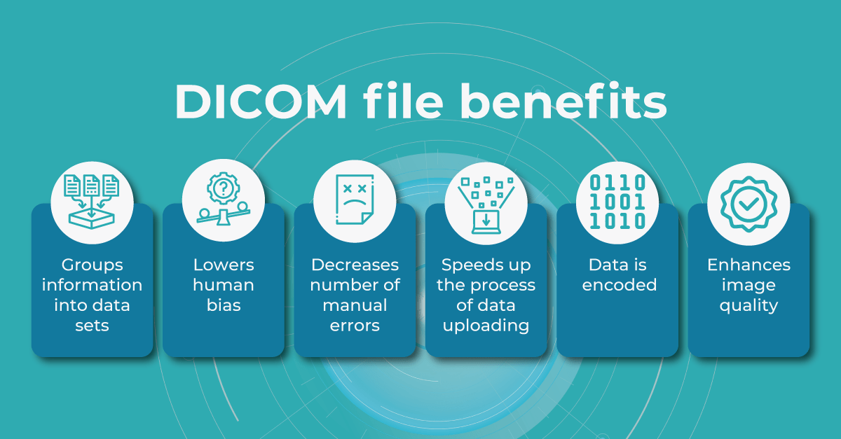
The DICOM standard ensures interoperability between different vendors’ OCT devices and facilitates seamless data sharing and analysis. The main difference between DICOM and other image formats is that it groups information into data sets. A DICOM file consists of several tags, all packed into a single file. It stores such info as:
- demographic details about the patient
- imaging study’s acquisition parameters
- image dimensions
- matrix size
- color space
- an array of additional non-intensity information necessary for accurate image display by computers.
If you have to enter the patient’s information manually, there’s always a chance you can misspell the name or other information. However, when using a DICOM file to store patients’ information and monitor patients’ health, eye care specialists can be sure the chance of human bias is much lower.
When you work in an optometry practice or a clinic, you may spend a lot of time filling in the details every time you upload a file. And if your clinic is busy and you do 30-50 uploads daily, it could take hours. Using DICOM image format significantly speeds up the process and reduces errors.

Another benefit of the DICOM image format is that the header data information is encoded within the file so that it cannot be accidentally separated from the image data.
DICOM files can be stored in a DICOM server or transmitted between DICOM-compliant systems using the DICOM network protocol (DICOM C-STORE or DICOMweb). DICOM SR (structure reporting) allows for the structured representation of measurement data and annotations in OCT images. It enables the storage of quantitative measurements, such as retinal thickness or optic nerve parameters, as structured data within the DICOM file.
In addition, eye care specialists are able to manipulate the brightness of the image when using the DICOM viewing software. Some areas of an image can be increased or decreased for a better viewing and diagnostic experience.
Is DICOM file format popular among OCT providers?
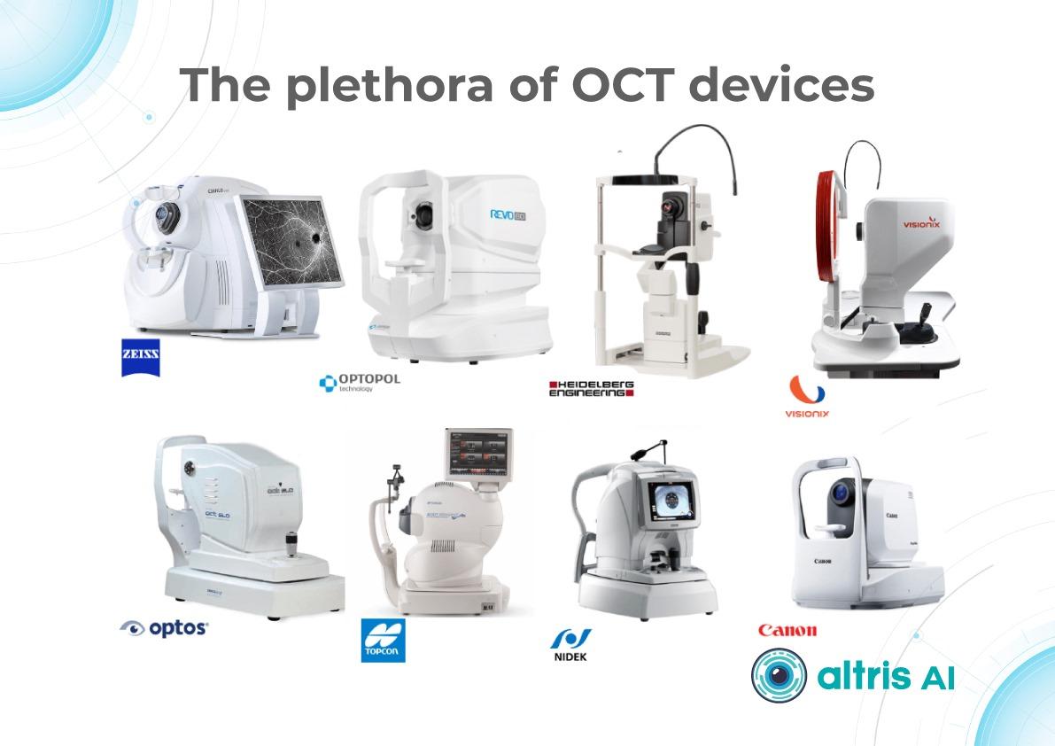
When it comes to optical coherence tomography, many OCT device manufacturers and software providers support the DICOM standard for storing and exchanging OCT images. Some of the prominent OCT providers that offer DICOM support include:
- Heidelberg Engineering is a well-known provider of OCT devices and software solutions for ophthalmology. They offer OCT devices like the Spectralis OCT, which supports DICOM connectivity. The DICOM capabilities of their systems enable seamless integration with PACS and other healthcare systems.
- Carl Zeiss Meditec is a leading manufacturer of ophthalmic devices, including OCT systems. Their OCT devices, such as the Cirrus OCT, are DICOM-compatible, allowing for efficient storage and sharing of OCT images with other DICOM-compliant systems.
- Topcon Medical Systems is another prominent provider of OCT devices. Their OCT systems, such as the Topcon 3D OCT, support DICOM connectivity, enabling interoperability with other DICOM-enabled devices and systems.
- NIDEK offers a range of ophthalmic imaging devices, including OCT systems. Their OCT platforms, such as the NIDEK RS-3000, support DICOM, allowing for seamless integration with DICOM-compliant infrastructure, such as PACS and EHR systems.
These are just a few examples of OCT providers that support the DICOM standard. It’s important to note that DICOM support may vary among different models and versions of OCT devices from each manufacturer. We recommend you consult with the specific manufacturer or review their product documentation to confirm the DICOM capabilities of their OCT systems.
Why do we recommend using DICOM file format with Altris AI?
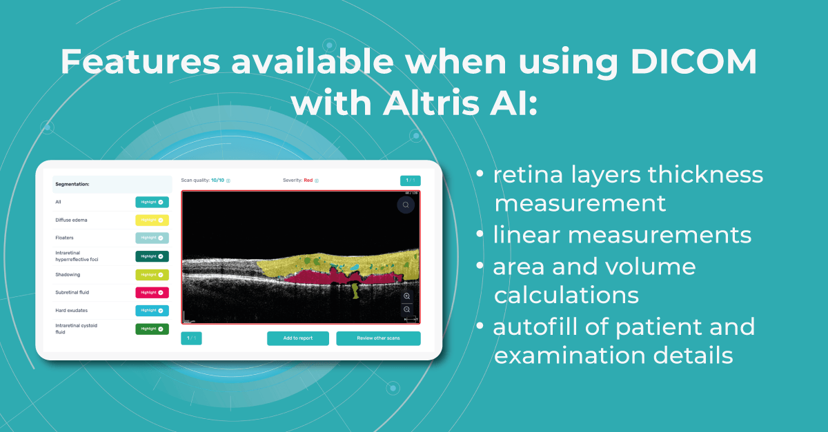
Modern DICOM viewer software extends beyond simple viewing. It can enhance image quality, generate additional data, take measurements, and more, and Altris AI is no exception. Using the DICOM image file gives you more opportunities within the platform.
Such features as
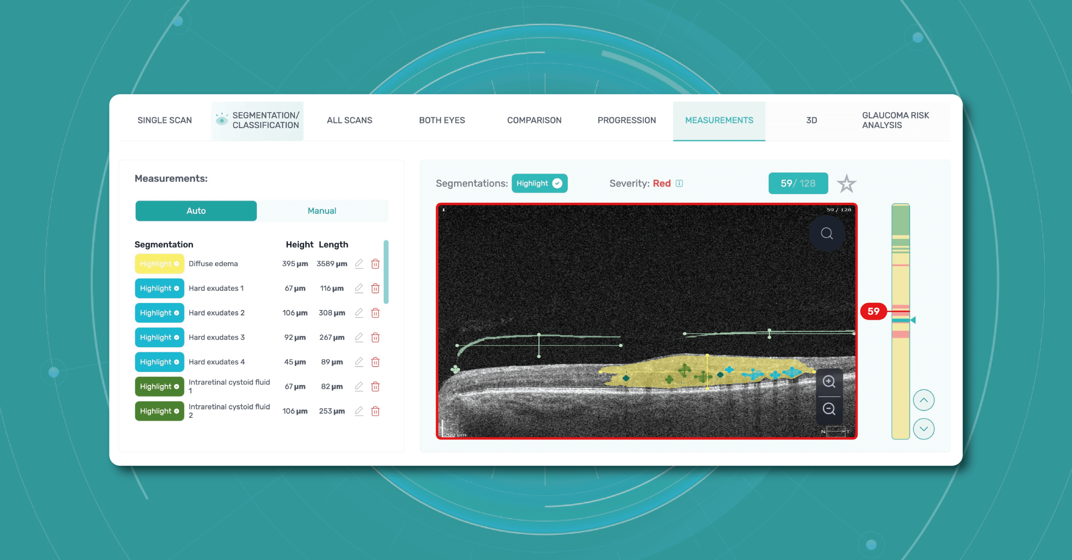
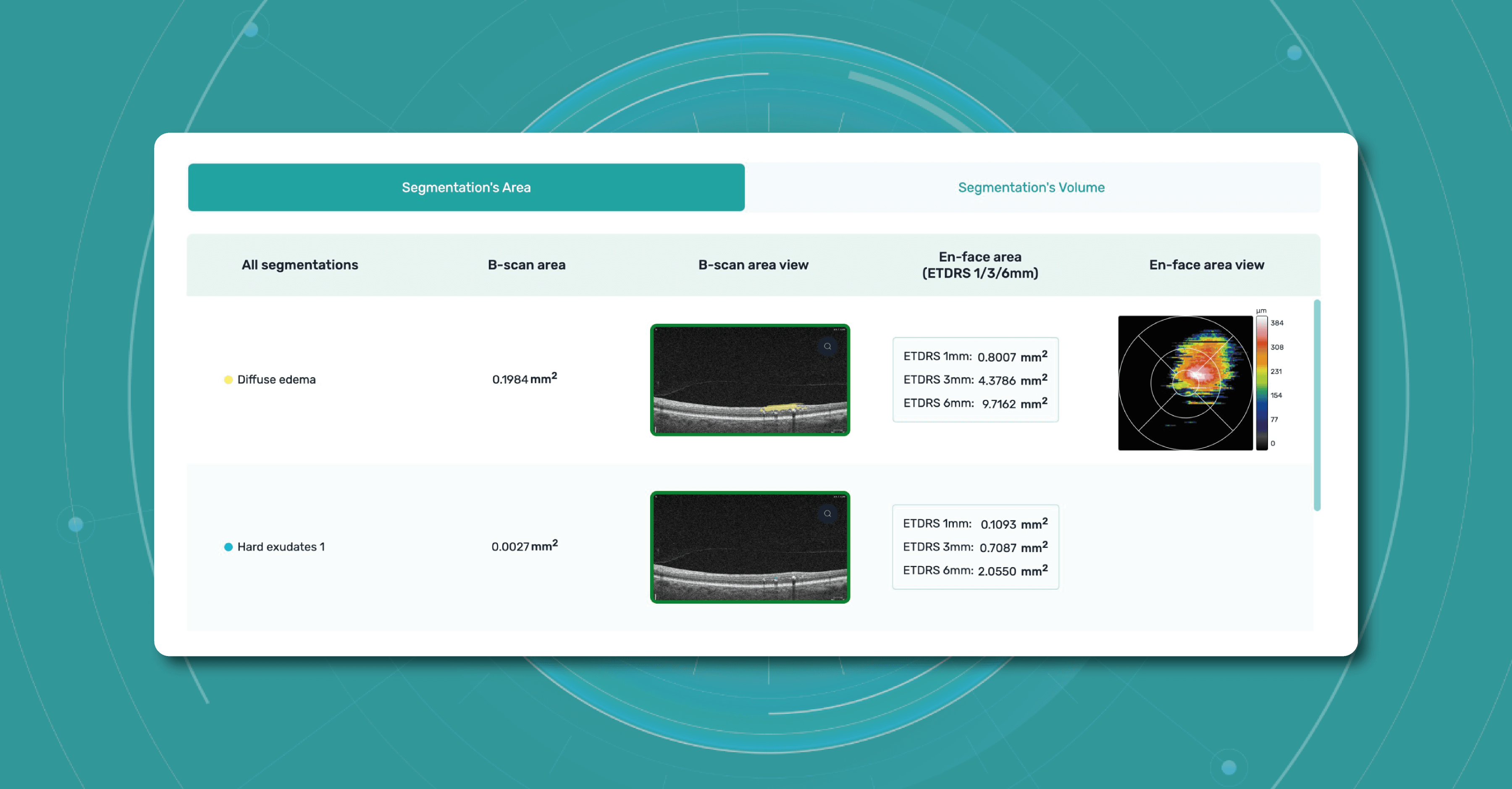
- retina layers thickness and linear measurements

- area and volume calculations

are only available when using the DICOM file format. This is because it contains the original image pixel data without modifying the study metadata. In case you upload an image, retina layers thickness won’t be available, as well as the measurements.
Another advantage of the DICOM format is that you can add patient and examination details in a few clicks by just uploading a DICOM file since this information is being pulled out automatically.

In the case of other image formats, when uploading an examination, you would have to manually fill in a bunch of information such as scan widths, eye type, etc.
Considering all mentioned above, using DICOM format files saves time, increases efficiency, and gives you more opportunities within the Altris AI platform.
Summing up
What is DICOM format? In conclusion, the DICOM file format proves to be a valuable asset for eye care specialists. Its unique characteristics, such as grouping information into data sets and incorporating standardized tags within a single file, offer many advantages.
This format ensures the preservation of accurate and comprehensive data, reducing the potential for human error and minimizing the risk of data loss or misinterpretation. The DICOM file format streamlines the archival, organization, and display of images, optimizing the workflow of eye care specialists.
By adhering to the DICOM standard, OCT devices and software solutions ensure compatibility, interoperability, and consistent data representation across different platforms. This enables efficient communication and collaboration among healthcare professionals, enhances research capabilities, and promotes the broader use and exchange of OCT imaging data.
With its widespread adoption and compatibility with various medical imaging systems, DICOM empowers ophthalmologists and optometrists to provide efficient and high-quality care while promoting seamless collaboration and knowledge sharing within the field. Ultimately, the DICOM file format plays a vital role in enhancing patient care, advancing research, and fostering innovation in the field of eye care.
-

How 7 Leading Optometry Centers Provide Innovations in Eye Care
 Maria Martynova
08.05.20239 min read
Maria Martynova
08.05.20239 min readDisclaimer: USA FDA 510(k) Class II; Altris Image Management System (Altris IMS); AI/ML models and components intended to use for research purposes, not for clinical diagnosis purposes.
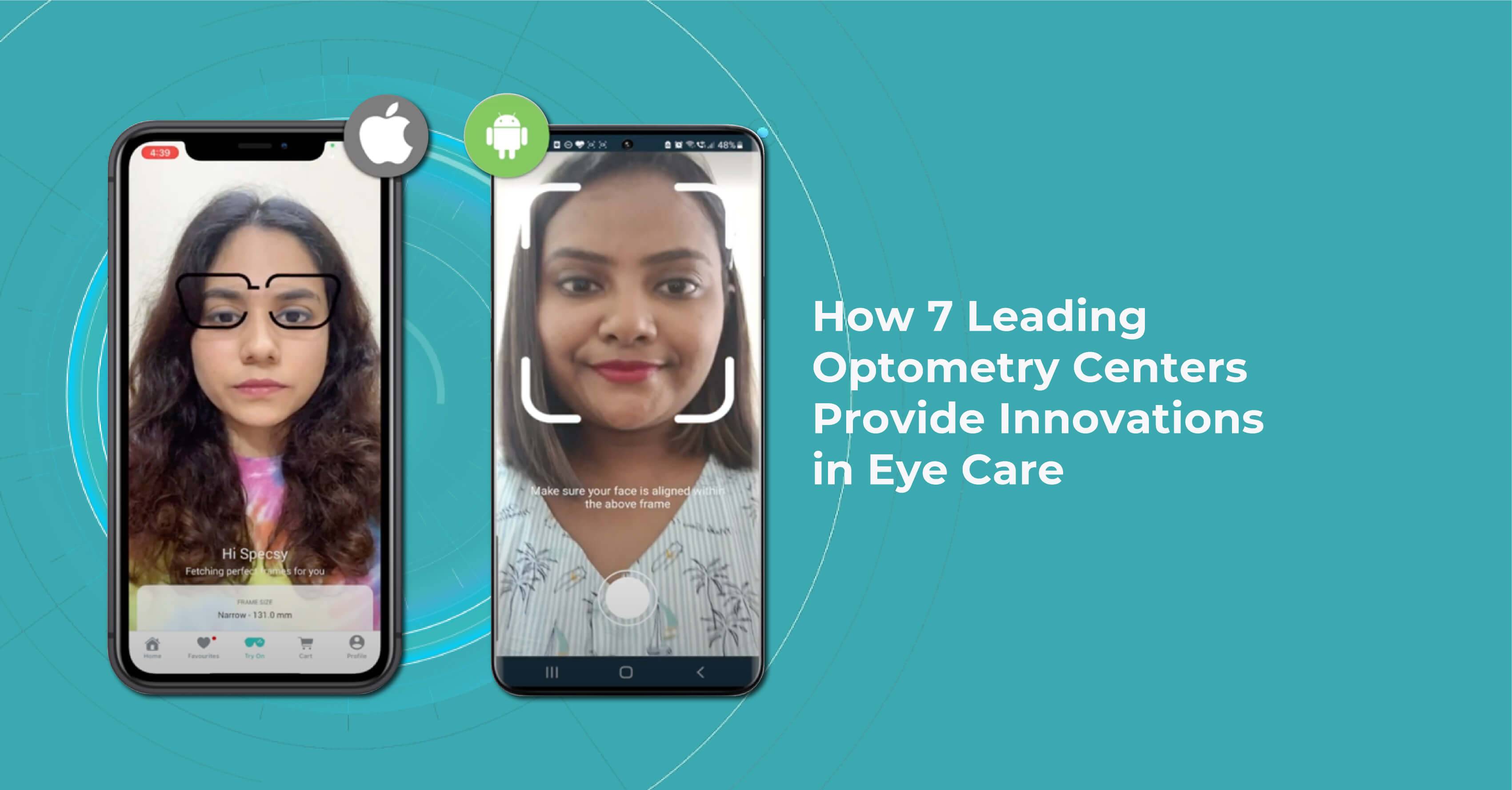
Top modern optometry centers are not afraid of embracing effective eye care innovation. Some offer home eye tests, others create mobile apps to try on frames remotely. There are optometry centers that use artificial intelligence to empower optometrists in OCT/ fundus interpretation. We’ve collected 7 optometry centers that are using technology now to win the competition.
From advanced diagnostic and treatment technologies to personalized care and patient education, these centers are transforming the way clients approach and bring innovations in eye care.
Optometry meets technology: AI, AR, mobile apps, and home eye tests
Augmented Reality (AR), mobile apps, and home eye tests are emerging trends that are changing the way people receive eye care.
- AR technology uses the camera lens on a mobile device or your PC as the method to deliver information and graphics. A user accesses an AR application, and the camera viewpoint incorporates the data directly into the perspective in real time. With AR apps for eyewear and exams, anyone can have a large selection of glasses and other services from their homes, offices, or on the go.
- Mobile apps offer a wide range of eye care services, from information on eye health and tips for maintaining healthy vision to virtual vision screenings. Moreover, mobile apps are also used to educate both young and experienced optometrists. We strongly believe that educational mobile apps inevitably become an additional efficient tool for OCT education because they are accessible and interactive.
- Another one of the innovations in eye care is Home eye tests are also often enabled by digital vision testing tools. They are becoming more and more common and offer a convenient and cost-effective way to monitor vision changes.
- As for AI use in optometry practice, it allows its users to see a broader perspective of a patient’s eye health. Incorporating AI streamlines billing procedures, expands the input of electronic health records (EHRs), optimizes claims management, and improves cash flow. AI technology can also be used in cooperation with AR assisting in the glasses selection.
Although these innovations in optometry and ophthalmology provide more comprehensive access to eye care and improve patient engagement, many optometry practices are still hesitating to add such innovations to their routine. That is why we prepared the info about 7 famous optometry practices that are already using innovations in eye care.
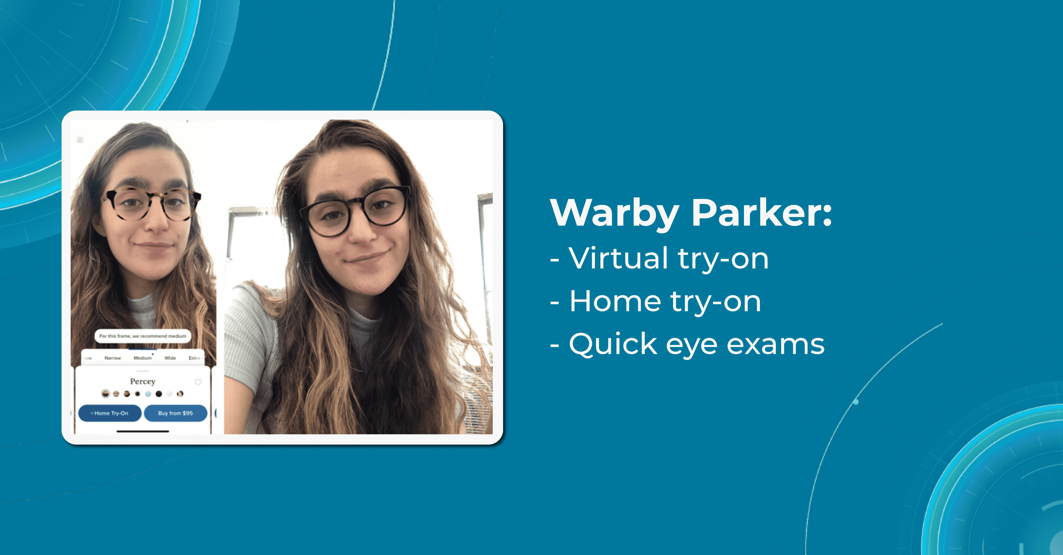
Warby Parker

Warby Parker started its way in 2010 when the founders of the company were students. One of them lost his glasses during a tourist trip. The cost of replacing them was so high that he spent his first semester of graduate school without them. That is why the company’s mission is to provide affordable, high-quality eyewear to consumers, while also addressing the issue of access to vision care.
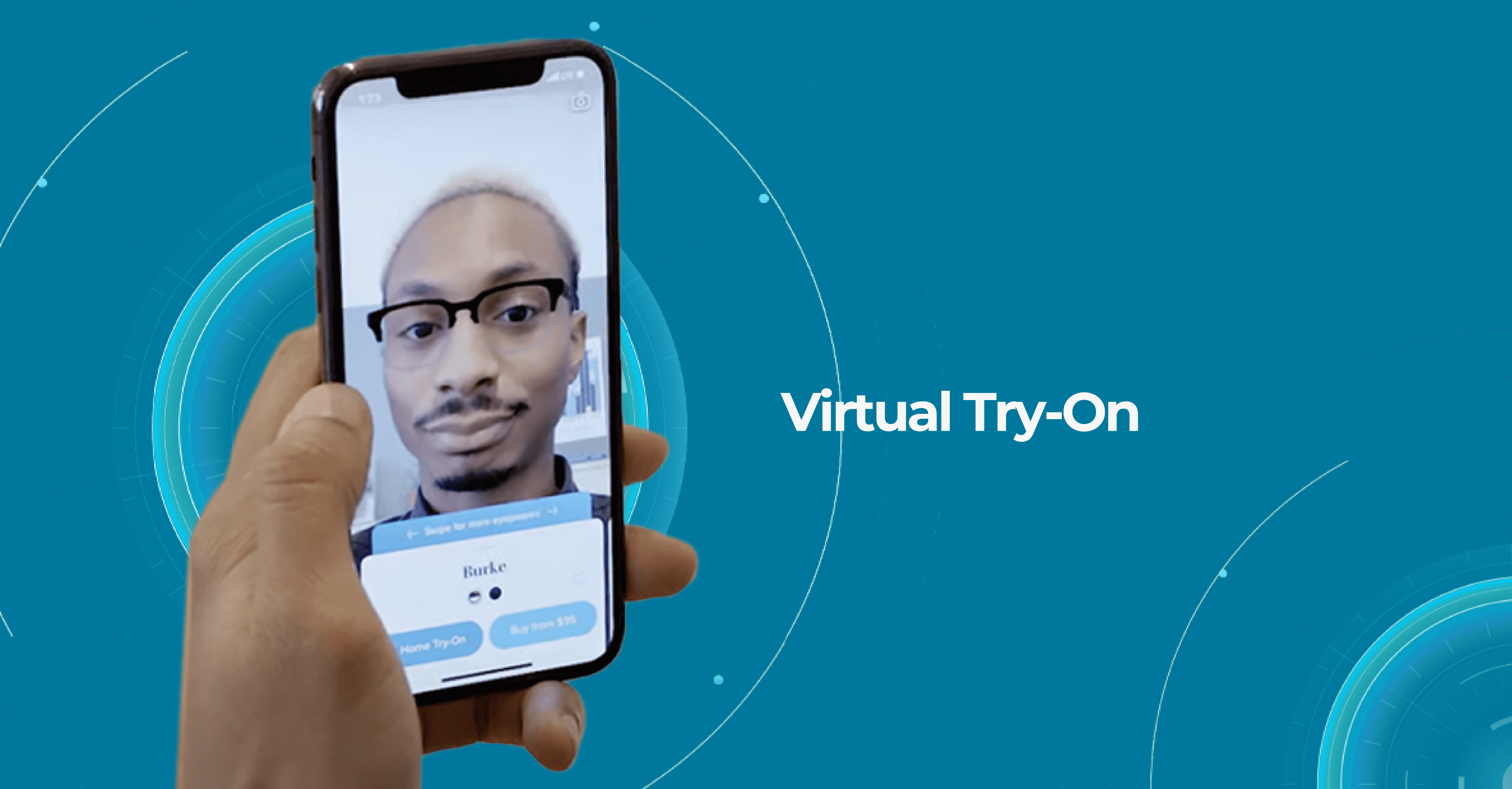
One of Warby Parker’s unique innovations in eye care is its Home Virtual Try-On program, which allows customers to try on up to five frames at home for free before making a purchase. This program makes it easier for customers to find the perfect pair of glasses and eliminates the need for them to go to a physical store to try on frames.

Warby Parker also offers an online eye exam called the Virtual Vision Test. It is designed to provide customers with a convenient and affordable way to obtain a prescription for glasses or contacts from the comfort of their own homes.
The Virtual Vision Test is a telemedicine service that uses technology to allow customers to take an eye exam using their computer or smartphone. The test is not meant to replace a comprehensive eye exam performed by an eye doctor, but rather to provide a convenient option for those who need a prescription renewal or have mild refractive errors.
After completing the test, the results are reviewed by a licensed ophthalmologist or optometrist, who will issue a prescription if appropriate. The customer can then use the prescription to purchase glasses or contacts from Warby Parker or any other provider.
Lenskart

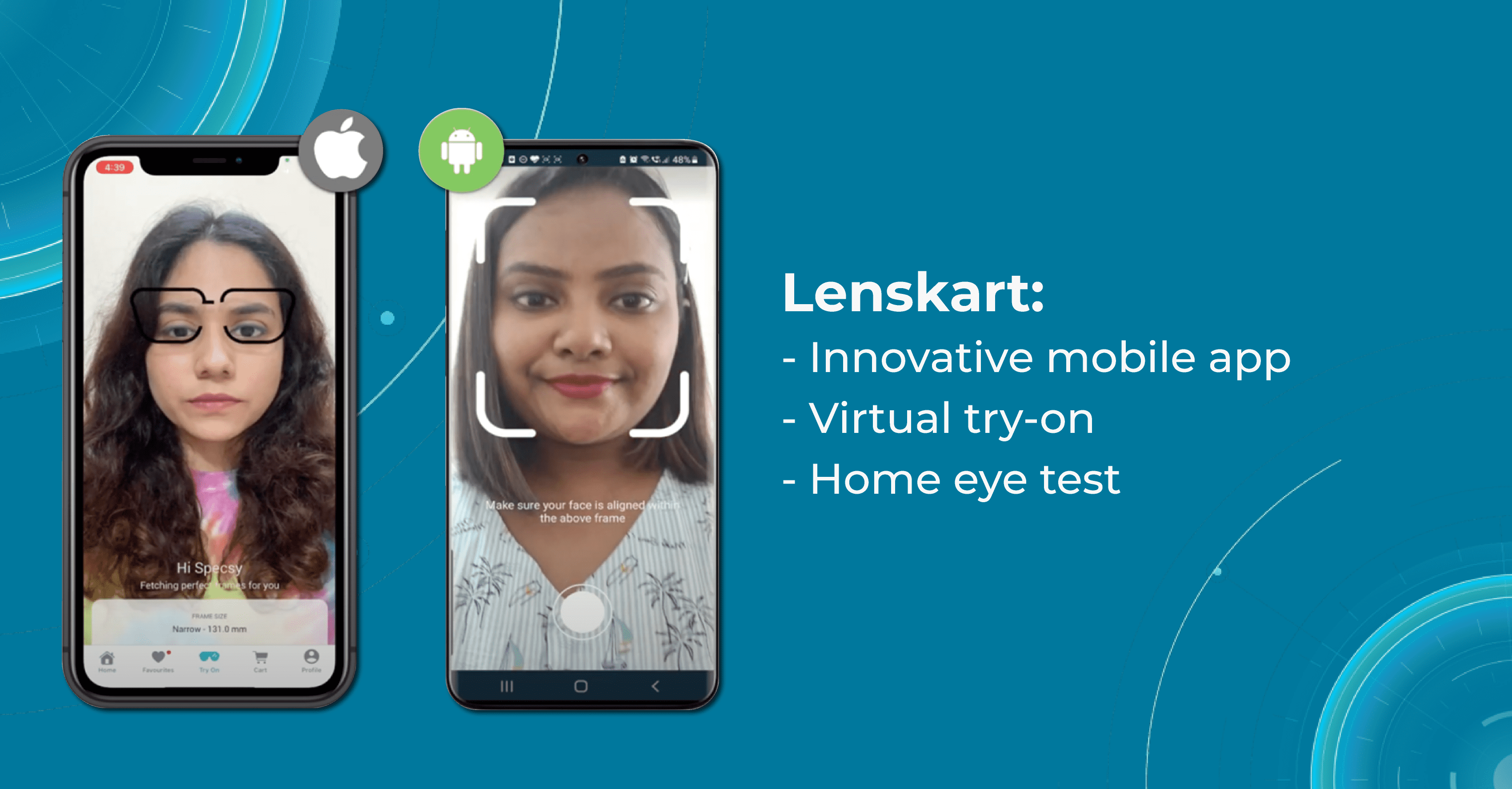
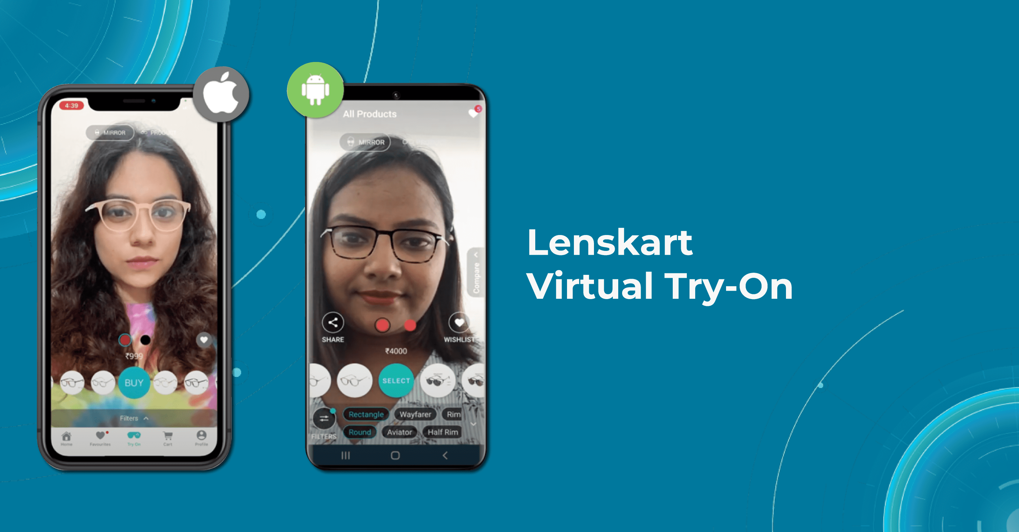
Lenskart is a fast-growing company of innovations in eye care in India focused on making eyewear more affordable for everyone. To achieve this goal, the company has developed a number of innovative technologies and business models, including a mobile app that allows customers to try on frames virtually and a home vision testing service that allows to check their prescriptions from the comfort of their own home.
One special feature of the Lenskart app is the “3D Try-On” feature, which uses 3D imaging technology to create a model of the customer’s face and allows them to try on different frames virtually. This feature helps get a better sense of how a particular frame will look on a customer’s face before making a purchase.

Another one of Lenskart’s innovations in eye care is the Home eye test, designed to provide people with a convenient and affordable way to obtain a prescription for glasses or contact lenses. To take the Lenskart Home Eye Test, customers must first book an appointment on the company’s website or mobile app.
The eye test includes a visual acuity test, a color vision test, and a refractive error test. The optometrist will also check the customer’s eye health and recommend any necessary follow-up exams or treatments. After the test, the optometrist will provide a prescription, which the customer can use to purchase glasses or contacts from Lenskart or any other provider.
SmartBuyGlasses

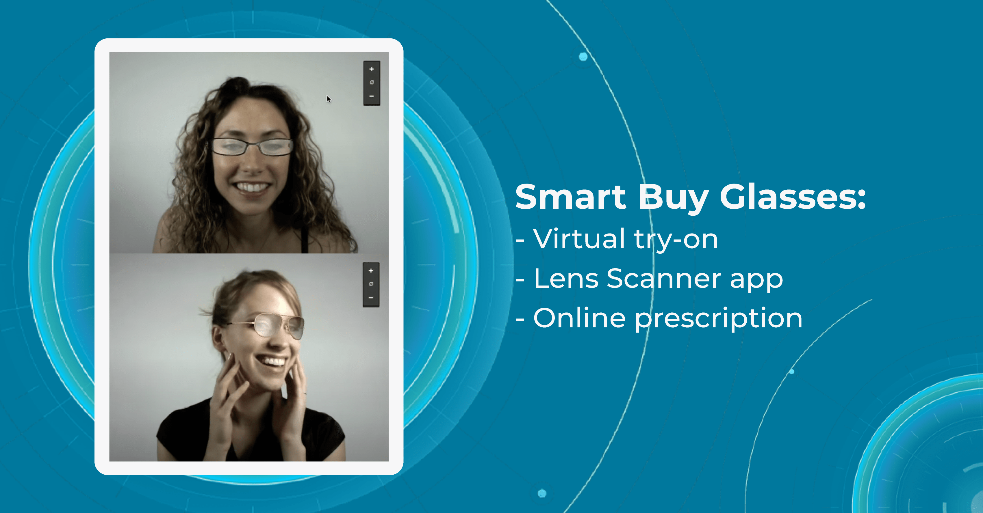
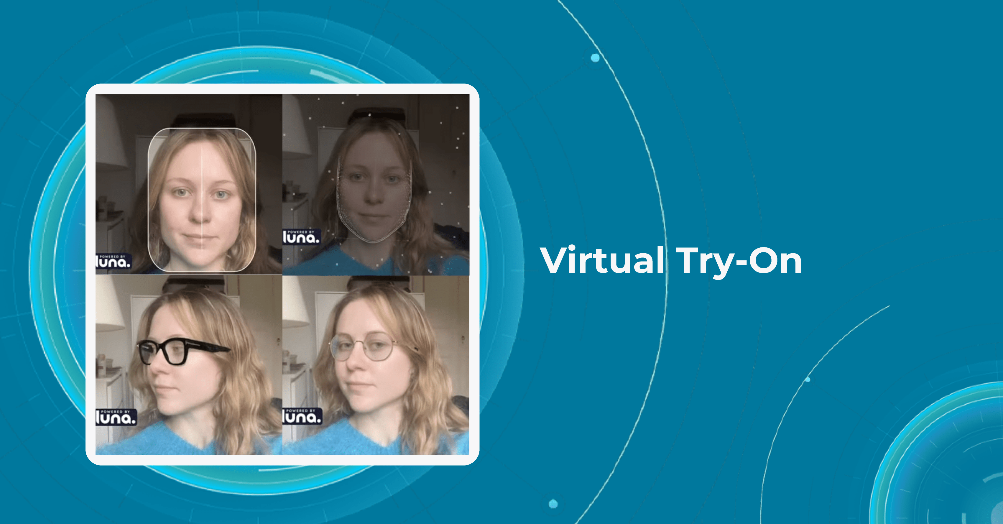
SmartBuyGlasses is an online eyewear retailer that was founded in 2006. The company is headquartered in Hong Kong, but it operates in more than 20 countries worldwide. Company’s Virtual Try-On feature is available on the website and allows customers to upload a photo of themselves and try on glasses virtually using augmented reality.
After the website generates a 3D model of the customer’s face, they can adjust the position and size of the glasses to get a better sense of how they will look on their faces. The virtual try-on innovations in eye care also allow to share images of themselves wearing the glasses with their friends and family to get feedback on which pair looks best on them.

Another eye care innovation of SmartBuyGlasses is a Lens scanner app that uses advanced technology to scan the user’s current eyeglasses lenses and analyze the prescription, allowing to order a new pair of glasses online without visiting an eye doctor.
The app works by instructing the user to place their current eyeglasses on a flat surface and position their smartphone camera above the lenses. The app then captures a series of images and uses advanced algorithms to analyze the curvature, thickness, and other factors of the lenses to determine the prescription.
GlassesUSA

GlassesUSA is an innovative and socially responsible eyewear retailer that is committed to providing quality products and services to its customers. With its focus on technology, sustainability, and social impact, GlassesUSA has become a popular choice for customers in the United States and around the world.
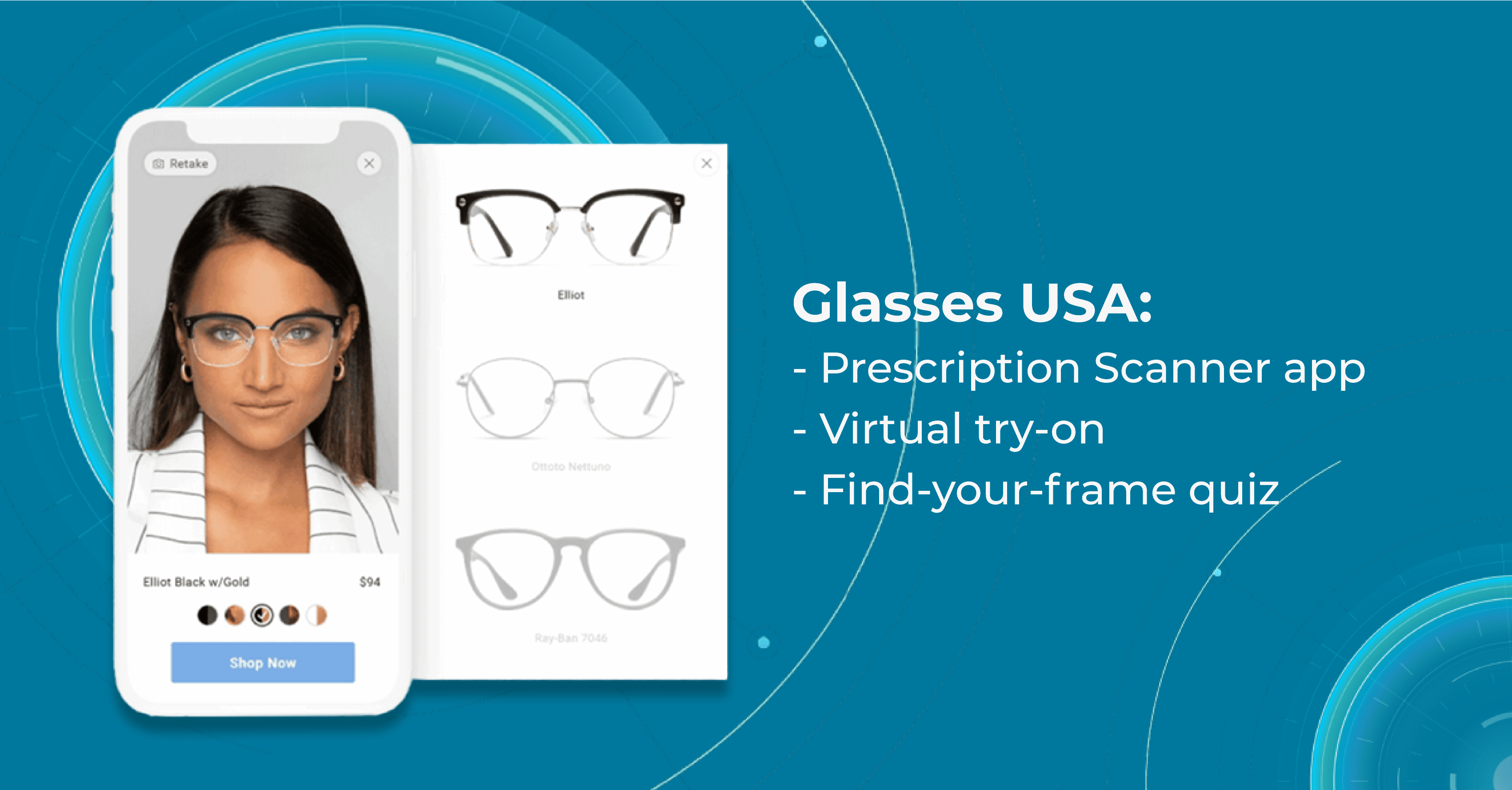
One of the innovations in eye care of GlassesUSA that is worth paying attention to is a Prescription Scanner app. The app works by guiding the user through a series of steps to scan their face and eyes using their smartphone camera. It uses advanced algorithms to analyze the user’s facial features and measure the distance between their pupils, which is a crucial factor in determining the correct prescription for eyeglasses.

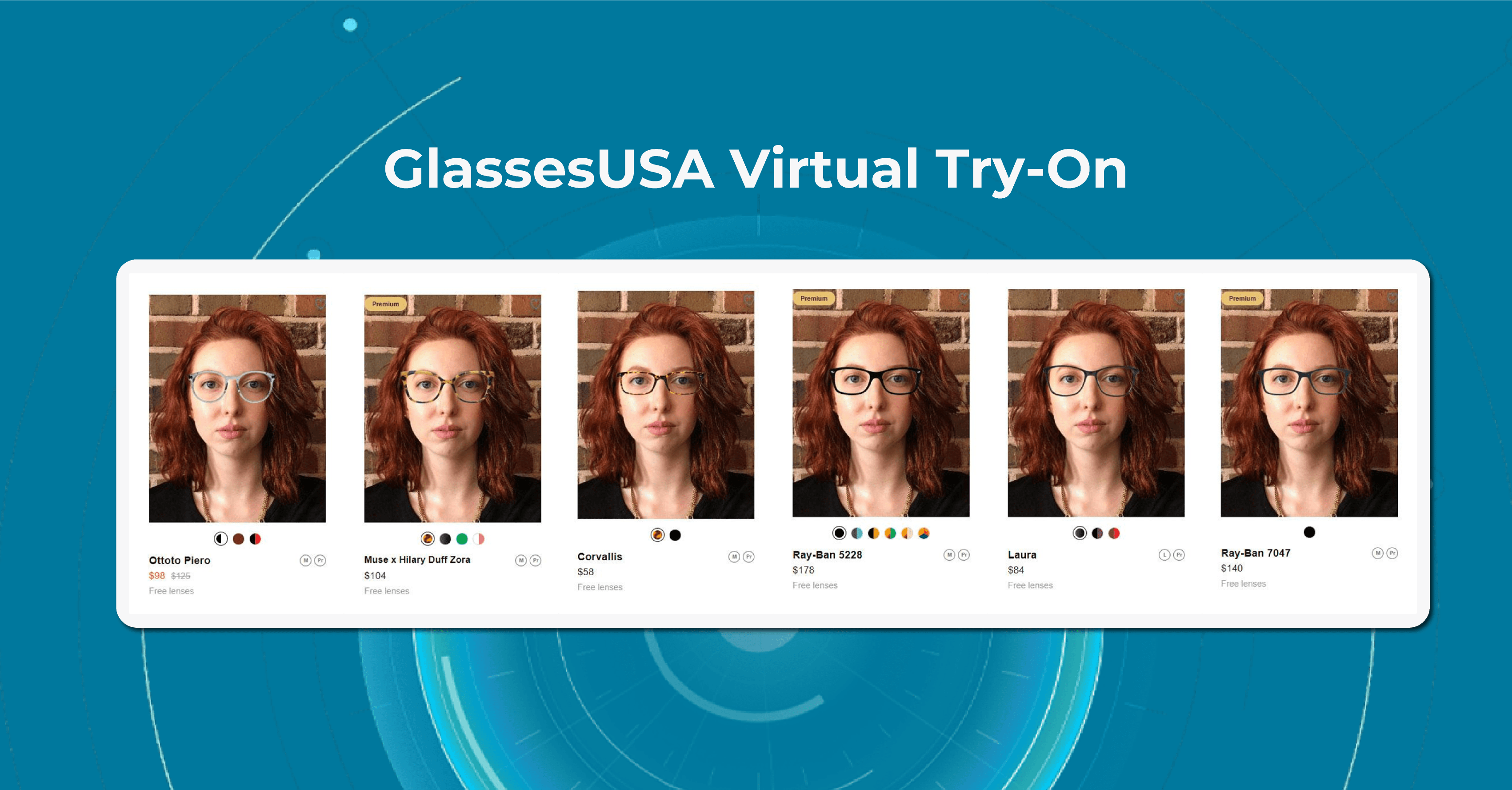
Once the scanning process is complete, the GlassesUSA app provides the user with their personalized prescription and recommendations. The app also offers a Virtual Try-On feature that allows users to see how different frames will look on their faces before making a purchase.
Another feature is a Find-your-Frame Quiz on the website. The quiz consists of a series of questions that ask users about their face shape, personal style, and preferences for eyeglass frames, such as color, material, and shape. Based on the user’s responses, the specially designed program generates a personalized selection of eyeglasses frames that are recommended for their face shape and style preferences.
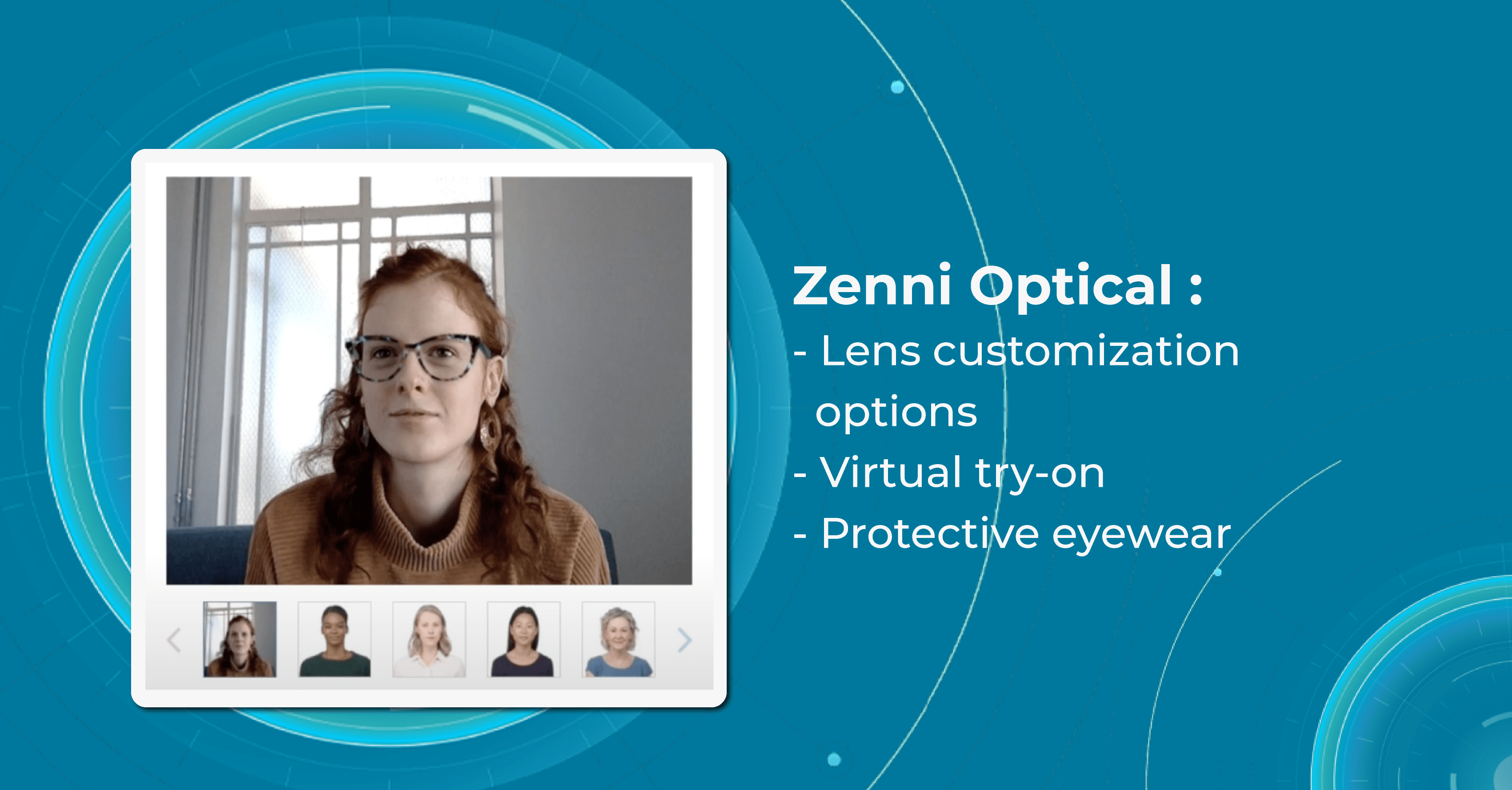
Zenni Optical

Zenni Optical offers a wide range of eyewear products, including prescription glasses, sunglasses, and sports eyewear. The company offers glasses at significantly lower prices than traditional brick-and-mortar stores, which has made it a popular choice for customers.
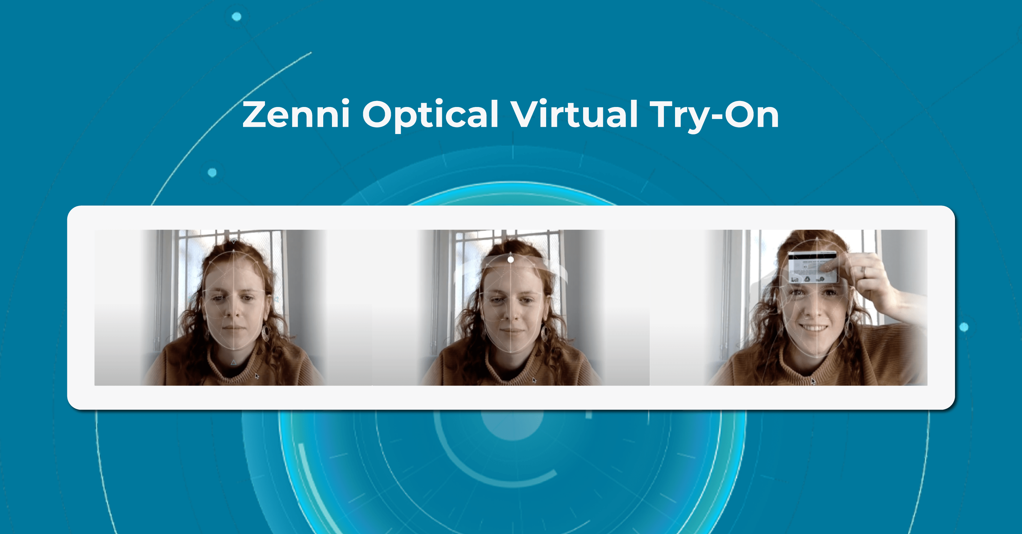
Company’s Virtual Try-On feature uses advanced AR technology to create a 3D model of the user’s face, allowing them to see how different frames will fit and look on them.

To use the Virtual Try-On innovations in eye care, users simply need to upload a photo of themselves or use their computer or smartphone camera to take a live video. This feature then maps the user’s facial features and displays a selection of eyeglasses frames that can be tried on virtually. Users can then select different frames to see how they look from different angles, and can even compare different frames side-by-side.
The Zenni Optical Virtual Try-On is a convenient and easy-to-use tool for anyone in the market for a new pair of glasses. It allows users to see how different frames will look on their faces without the need to visit a physical store or try on multiple pairs of glasses.
VSP Global

VSP Global is a leading eyewear company that was founded in 1955 by a group of optometrists who wanted to provide affordable eye care. Today, VSP Global is a major player in the optometric industry and offers its customers a wide range of services and products.
The company works with a network of over 40,000 eye doctors and optometrists to provide affordable and accessible eye care to its customers. VSP Global also offers other eye care services, such as telehealth consultations, on-site eye exams for businesses and schools, and a mobile eye clinic that serves underserved communities.

AI for OCT
FDA-cleared system for your business
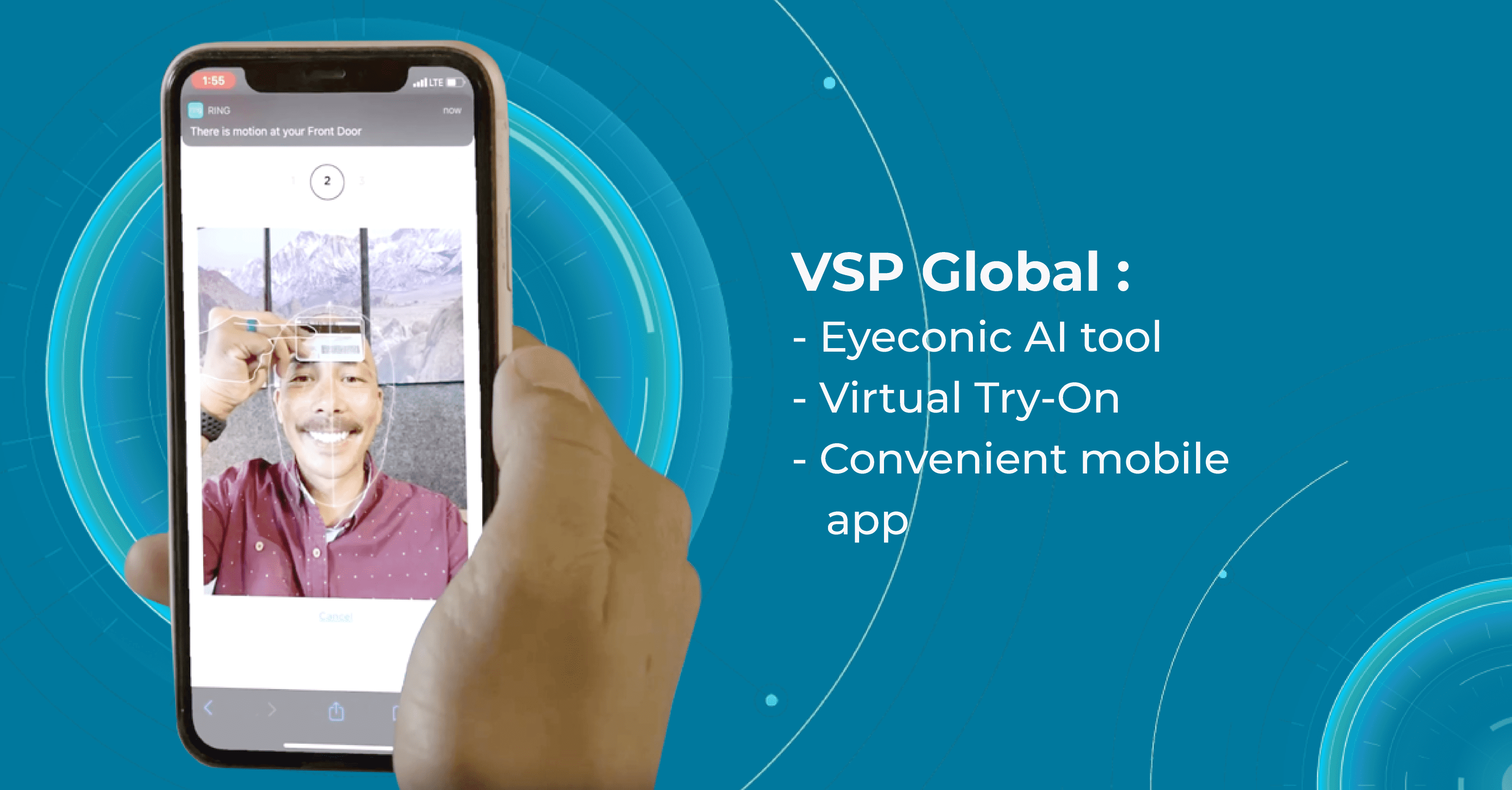
And as every company from this article, VSP Global has a strong focus on technology and innovations in eye care. The company has developed a number of proprietary technologies, including an AI-powered platform called Eyeconic that helps customers find the right eyewear.
Eyeconic uses machine learning algorithms to analyze a customer’s facial features and suggest frames that would fit their face shape and size. VSP Global has also developed a mobile app called myVSP that allows customers to manage their vision benefits, find an eye doctor, and order contact lenses online.
iSight+

Another AI-oriented optometry center is iSight+, located in Hong Kong. iSight+ is an excellent example of how an optometric eye care center didn’t hesitate and chose to provide innovations in eye care and a more in-depth examination of the macula.
Andy Meau. Optometrist, the owner of ISight+ Optometric Eye Care center:
“Altris AI will be a great tool in helping to monitor patients with existing macular diseases. I am also honored to be the first EPC in Hong Kong to provide this service.”
In addition, the eye care center is also equipped with advanced optometric technologies, digital photography systems, and optical coherence tomography (OCT), which helps to provide the highest quality eye examination.
Summing Up
Optometry centers can significantly benefit from incorporating innovations in eye care, such as augmented reality, artificial intelligence, and mobile apps, into their practice. These technologies enhance the patient experience, improve diagnostic accuracy, and streamline clinical workflows.
Moreover, the use of innovative technology can help optometry centers stay competitive in a rapidly evolving healthcare landscape. Patients are increasingly tech-savvy and expect healthcare providers to offer convenient, digital solutions that meet their needs. By embracing innovative technologies, optometry centers can attract new patients and retain existing ones, while also increasing operational efficiency and reducing costs.
Of course, there may be concerns about the cost and complexity of integrating new technologies into an optometry practice. However, the benefits of doing so can far outweigh these potential challenges. With careful planning and implementation, optometry centers can successfully leverage AR, AI, and other innovations in eye care to enhance patient care, improve clinical outcomes, and thrive in a rapidly changing healthcare environment.
-

Future of Optometry: How will Optometry Practice Look in 2040?
 Maria Znamenska
29.03.20239 min read
Maria Znamenska
29.03.20239 min readFuture of Optometry: How will Optometry Practice Look in 2040?
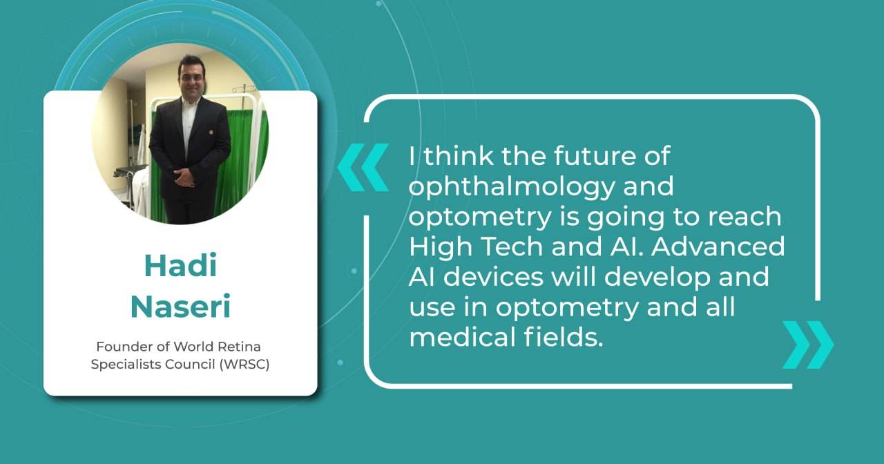
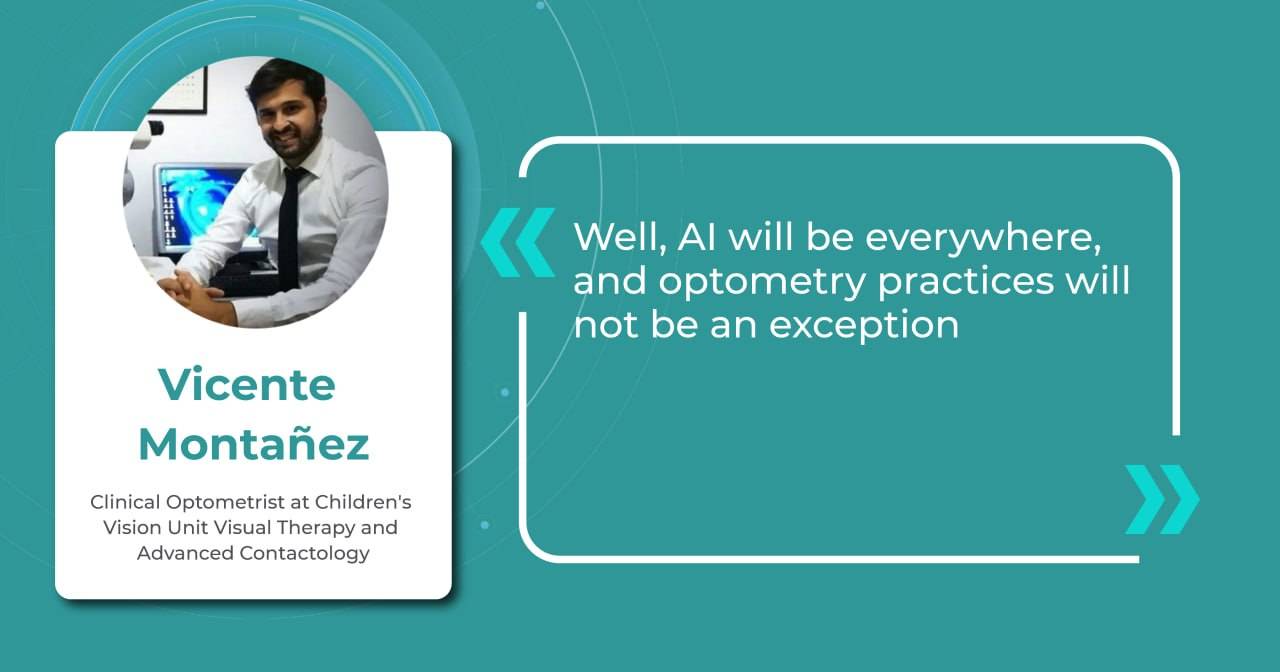
In the next two decades, we can expect to see a paradigm shift in the way optometry is practiced. Advances in new technology, such as AI (artificial intelligence), machine learning, and virtual and augmented reality, are expected to revolutionize how optometrists diagnose, manage, and treat eye-related problems. Optometry’s future is promising for those who are ready to embrace innovations.
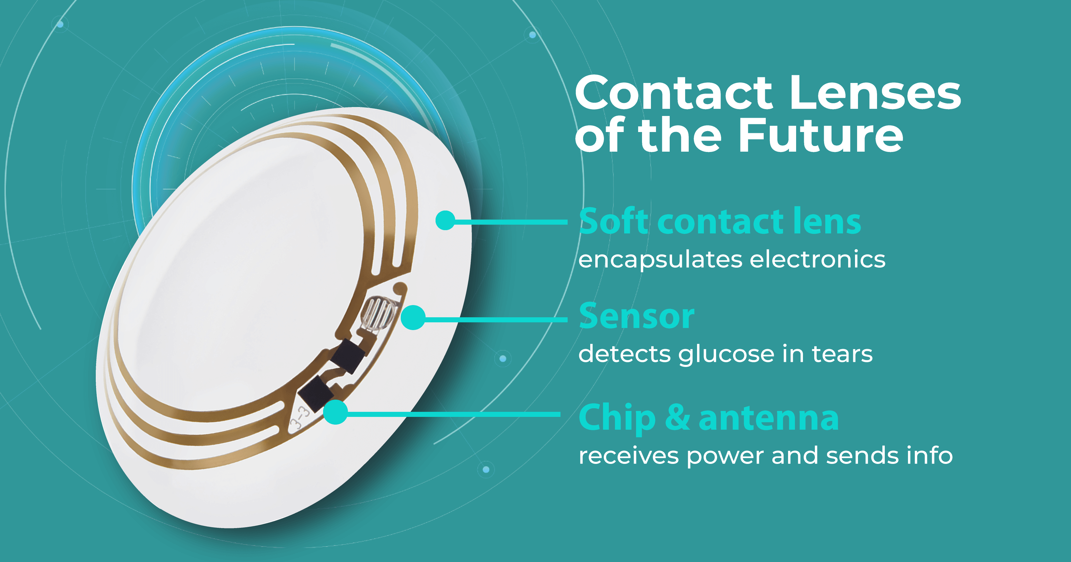
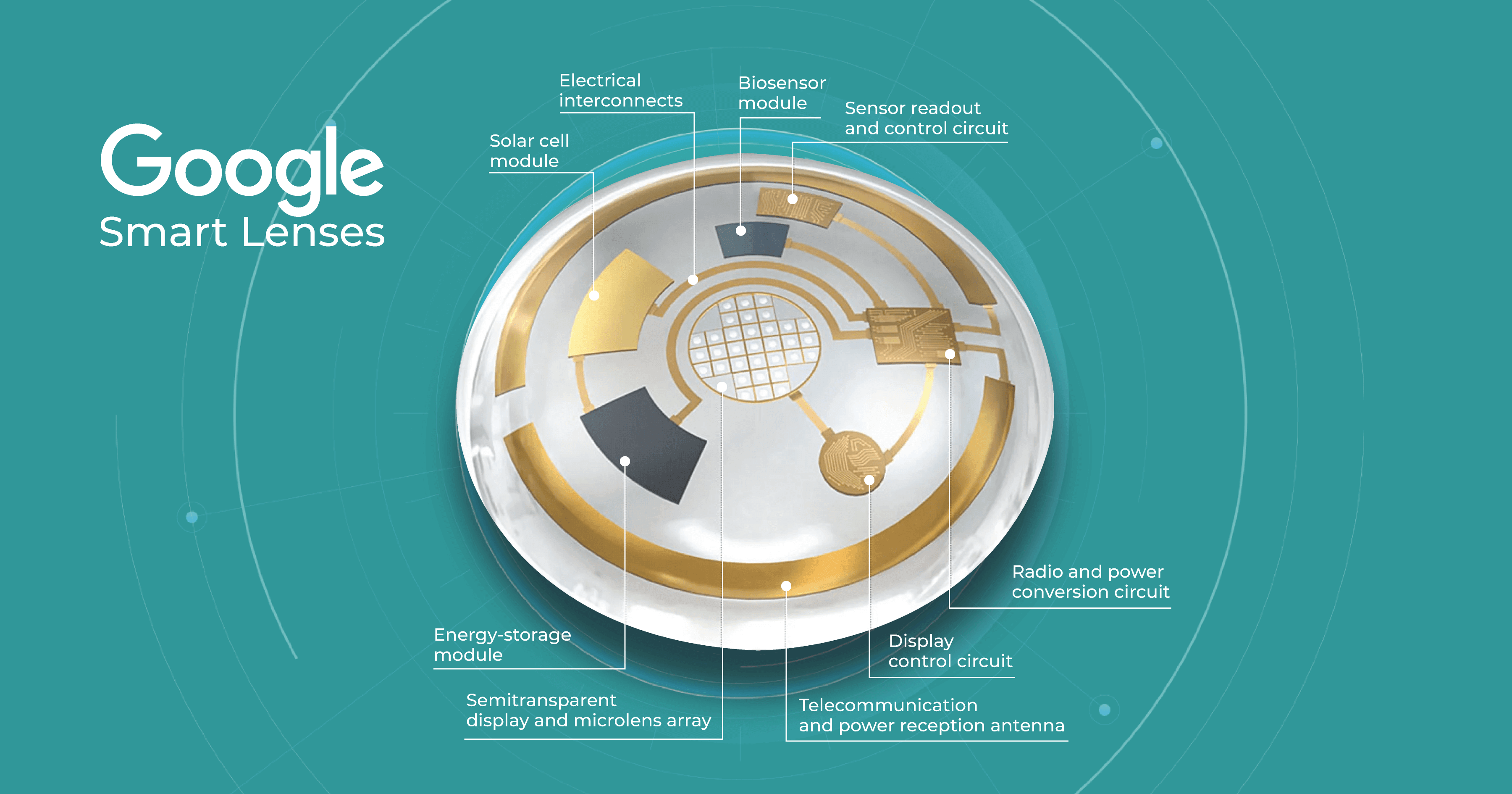
For example, smart contact lenses that can monitor blood sugar levels for diabetic patients or detect early signs of glaucoma are already in development, and they could become mainstream within the next 20 years.

In addition to the innovations, changes in demographics will also play a significant role in shaping the future of optometry. The aging population will require more specialized eye care, particularly for conditions such as macular degeneration and cataracts, which are more prevalent in older adults. The rise of chronic diseases such as diabetes will also increase the demand for optometric services, especially in developing countries where access to healthcare is limited.
The future of optometry is exciting and holds great promise for patients and practitioners alike. In this article, we will explore some of the potential changes that ODs may face in the coming years based on the survey that we have conducted.
In the next 20 years, the technology in eye care will be represented by AI and is expected to revolutionize the field in several areas. Here are some ways AI is helping in optometry:
- Diagnosis and treatment. AI algorithms can analyze large amounts of patient data and provide accurate and fast diagnoses of eye diseases such as glaucoma, diabetic retinopathy, and age-related macular degeneration. AI could also help in designing personalized treatment plans for individual patients.
- Screening and monitoring. AI can help specialists screen patients for eye diseases more accurately and quickly. For example, a patient could take a picture of their eyes with their smartphone, and an AI algorithm could analyze the image for signs of eye disease. AI could also help monitor the progression of eye diseases over time.

- Enhance patient care. AI-powered tools could help ODs provide more personalized and comprehensive care to their patients. For example, the AI algorithm helps to select the most suitable eyeglasses or contact lenses for a patient based on their unique vision needs and lifestyle factors.
- Research and development. AI could help optometrists develop new treatments for eye diseases. By analyzing large amounts of patient data, AI algorithms could identify new patterns and potential treatments for eye diseases. Enhanced by AI precision, this enables more accurate identification and quantification of biomarkers, leading to better patient stratification, treatment monitoring, and prediction of therapeutic responses.
In addition, the implementation of AI can present various prospects for improving clinic operations, simplifying billing procedures, accelerating the input of EHRs (electronic health records), optimizing claims management, and boosting cash flow. As high-deductible health plans (HDHPs) gain popularity among employers and patients, revenue cycle management can be seamlessly integrated with AI, considering the increasing number of patients defaulting on their medical bill payments.

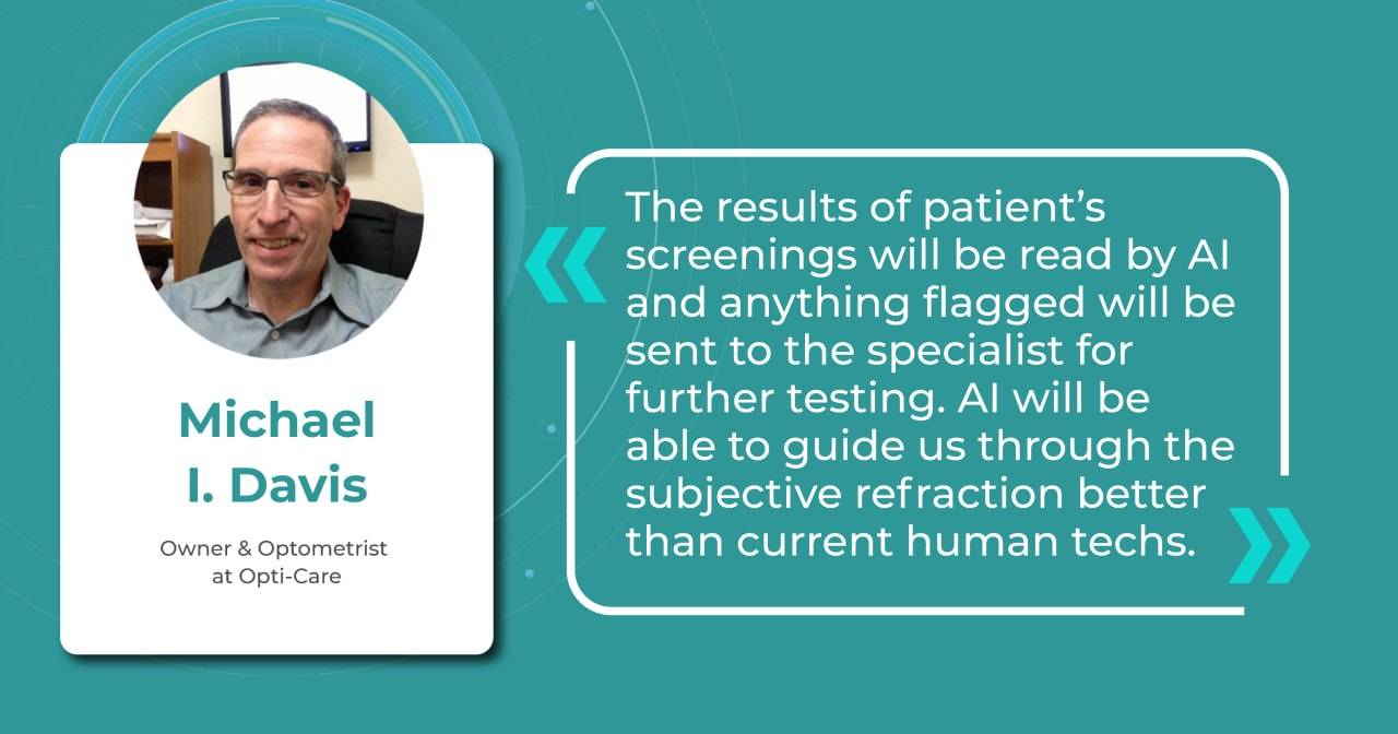
Although artificial intelligence is about to bring significant changes to the industry, it is important to remember that its effectiveness is limited to tasks that it has been specifically trained to perform. In contrast, AI may not perform well in areas outside its training.
Therefore, it is crucial to focus on enhancing ODs’ proficiency in utilizing AI instead of worrying about the possibility of job replacement. The integration of AI provides specialists with an opportunity to enhance patient outcomes on a global scale.
To utilize cutting-edge technologies proficiently, OD specialists must possess critical thinking skills and the ability to manage complex cases in real-time. Additionally, communication skills are essential, including cultural sensitivity, multilingualism, and familiarity with alternative communication platforms such as smartphone-based applications. These skills will be particularly important for optometry specialists in 2040.

Overall, AI has the potential to greatly improve the accuracy and speed of diagnosing and treating eye diseases, leading to better patient outcomes and a more efficient healthcare system.The evolution of OD and MD roles
In 2019, Richard C. Edlow, OD, claimed that nearly 20 million more routine and medical eye exams will be required in 2025 compared to 2015. That is the future of optometry that may look frightening because of the burden. The volume of surgery required for the aging US population will also increase. What is more, the number of cataract surgical procedures will also significantly increase—from 3.6 million in 2015 to 5 million in 2025. Add here the fact that the number of ophthalmologists will increase by only 2.1% in this same period.
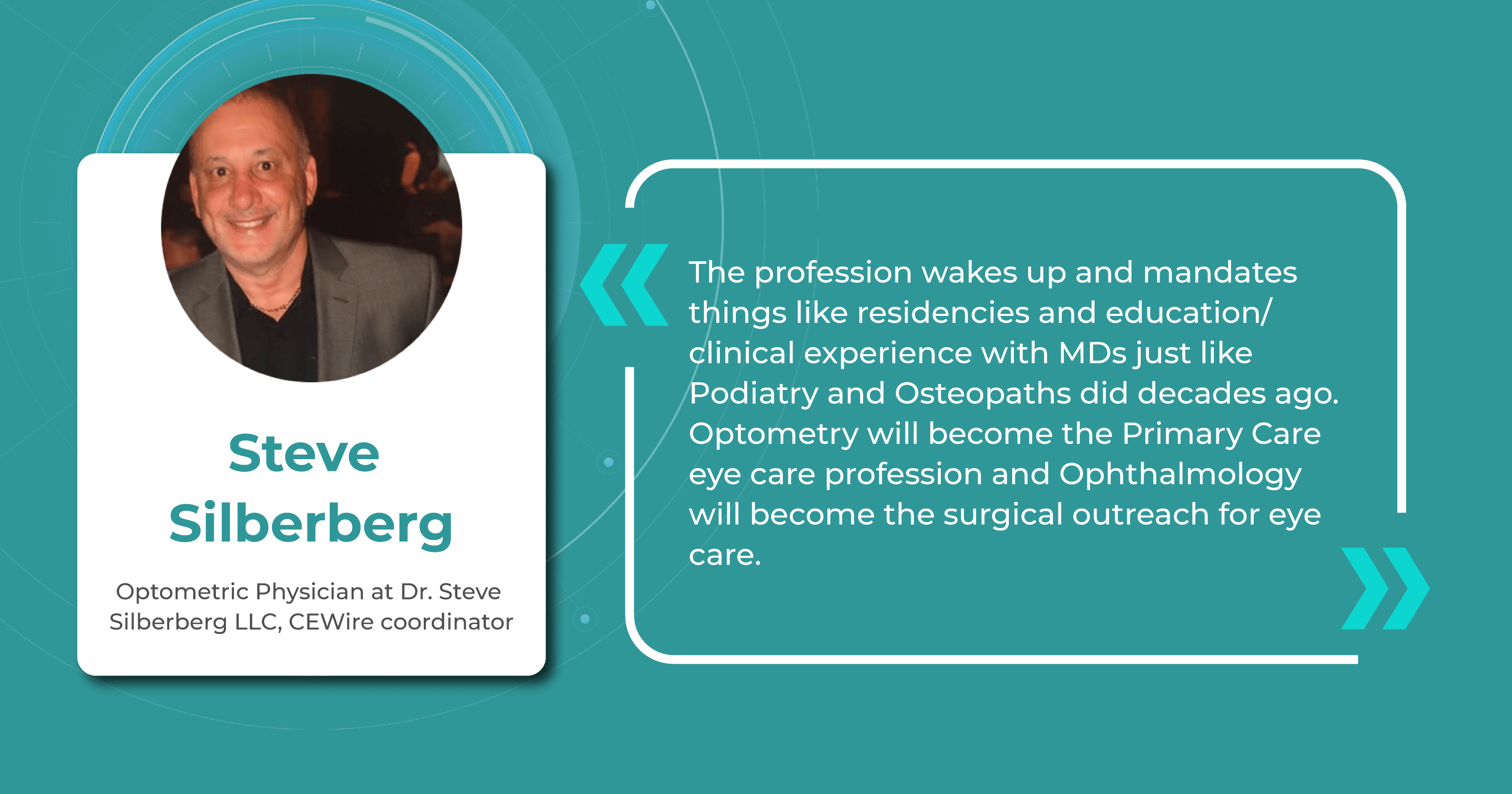
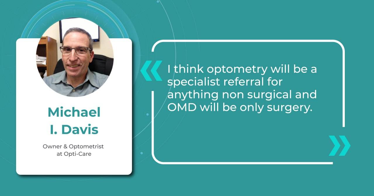
Given these facts, in the not-too-distant future, ophthalmologists will need to focus on surgical procedures, while optometrists will provide more medical care.

The field of ophthalmology must be fully prepared to meet the huge and growing demand for surgical procedures and therapeutic intravitreal injections. This brings us to the fact that the field of optometry, in turn, must be ready to manage the ever-increasing demand for medical ophthalmic services.
The roles of OD and MD are changing. With the advent of electronic healthcare, ophthalmologists are already spending more time on the computer than providing proper patient care. The ability to use innovative technology as well as in ophthalmology, digital thought processes, and critical thinking will create new opportunities in eye care as optometrists move further towards ‘data analysis’ and away from ‘data collection.’ OD specialists must ensure that they are properly trained in new technology in optometry and its advances to enhance, not inhibit, the quality of patient care.

It is also worth mentioning that despite the speed of new technology in optometry, the human relationship between patient and doctor remains the most powerful tool. To properly care for patients, ODs will need more than clinical skills, knowledge, or the latest technological advances. Patients need thoughtful, professional, kind, trusting, understanding, and caring optometrists.
As technology for the eye care advances, its education will also change. There may be more need for data analysis, less need for data collection, and an increased need for interpersonal skills (such as empathy, compassion, and bedside manner).
Future of Optometry: AI for OCT technology in optometry
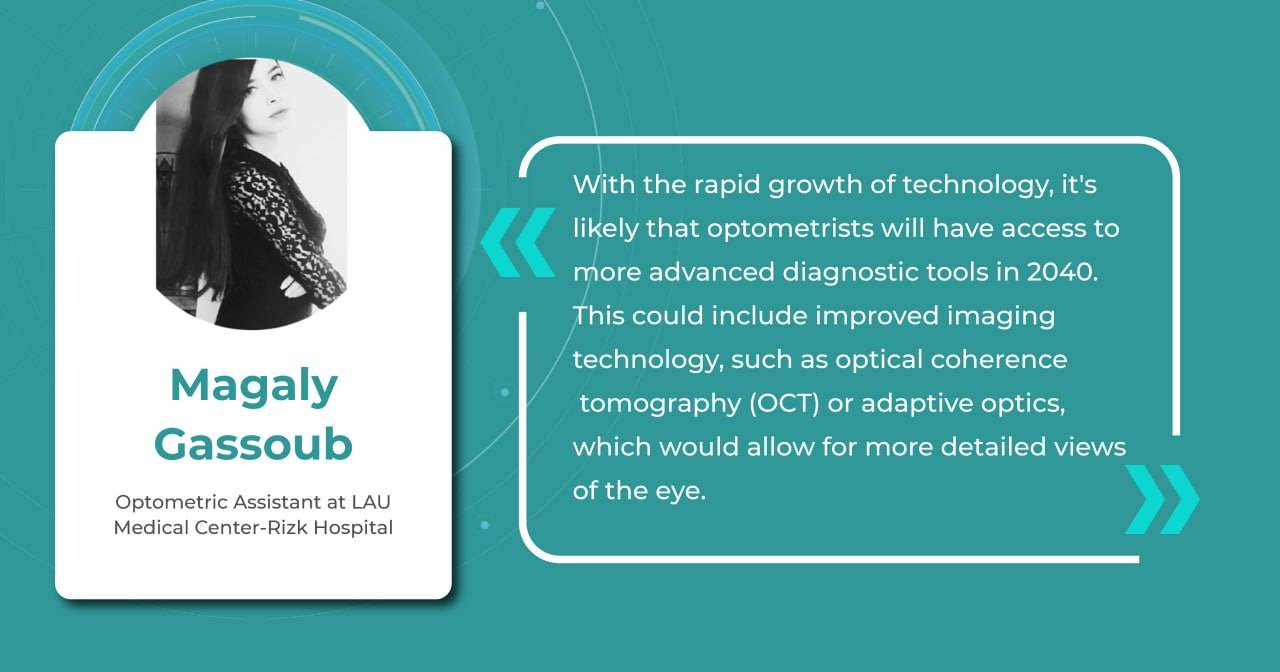
OCT has become an important diagnostic tool for the detection and treatment of various eye diseases, such as glaucoma, macular degeneration, and diabetic retinopathy. Its ability to obtain high-resolution cross-sectional images of the retina and optic nerve will broaden the horizons of technology and help optometrists detect and track changes in ocular structures that may not be visible during normal eye examination.

Here are some ways in which practitioners will benefit from implementing technology in the eye care:
- Improved diagnosis. OCT provides highly detailed images of the eye’s structures, allowing ODs to detect and diagnose eye conditions much earlier than with traditional methods. In fact, OCT is also called an optical retinal biopsy. This method makes it possible to examine 18 zones of the retina and detect minor or rare pathologies. This enables optometrists to provide timely treatment and prevent further damage to the eye.
- Better management of eye diseases is the future of optometry. OCT allows optometrists to monitor the progression of eye diseases such as glaucoma, ARMD, and diabetic retinopathy by taking detailed retinal images. It helps to determine the severity and stage of the disease, compare images after examination with documented results, and track disease progression. Moreover, with OCT examinations, ODs can also monitor the same patient to choose the most accurate diagnosis.
- Enhanced patient care. OCT is a noninvasive and painless procedure that is easy for patients to undergo. It uses safe laser light, avoiding all the side effects or risks. As the procedure is comfortable and effortless for both the ODs and patients, it helps to build stronger relationships by providing a less intimidating experience than other examinations.
- Increased revenue. Optometrists who offer OCT in their practices can generate an additional revenue stream by charging for the procedure and using it to attract new patients.
AI technology in optometry is improving diagnostic accuracy and enhancing practices’ overall efficiency. By automating tasks such as image analysis and data entry, AI frees up optometrists’ time, allowing them to focus more on patient interaction and complex decision-making. This streamlined workflow not only benefits practitioners but also improves the patient experience, making integration of AI into optometric practice not just a possibility but a new standard.
The future of Optometry: Focusing on myopia management
According to a survey conducted by the American Optometrists Association, nearly 70% of optometrists reported an increase in patient requests for myopia treatment in the last two years. Myopia is a rapidly growing problem worldwide. Only in the USA, it is predicted that by 2050 the number of patients will increase to 49.8%. As unfortunate as it may be, such a global epidemic of myopia will undoubtedly create an opportunity to expand the practice of specialized treatment.

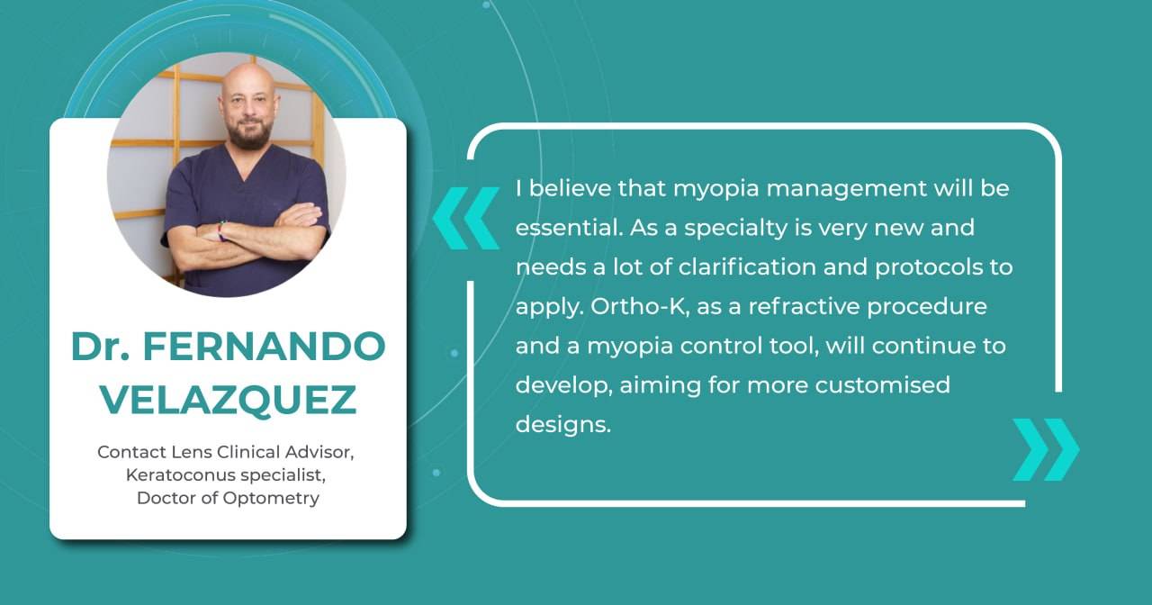
In the future, optometrists may manage myopia using a combination of approaches, and one of the most discussed is orthokeratology (ortho-K). This non-surgical approach that involves wearing specially designed contact lenses has been used to reduce the degree of myopia since the 1960s. Although this method is not new in optometry practice, many companies are still working hard to create new approaches and upgrade them. For example, two years ago, Johnson & Johnson Vision announced FDA approval of its Acuvue Abiliti Overnight Therapeutic Lenses for the management of myopia. That same year, CooperVision announced that its Procornea DreamLite night lenses for ortho-k had received the CE Mark from European regulators for slowing the progression of myopia in children and young adults.
Overall, the future of myopia management with new technology in optometry will likely involve a personalized, multi-faceted approach that combines various strategies to reduce the progression of myopia and improve vision.
Game-changing contact lenses
Research published in Advanced Materials Technologies claimed that contact lens sensors can be used to monitor many common diseases in the near future. The fact is that biomarkers in the lacrimal fluid make it possible to create diagnostic contact lenses. Such lenses would analyze these biomarkers and detect and treat systemic and ocular diseases such as diabetes, cancer, and dry eye syndrome.
It is predicted that in the near future, lenses will be able to monitor intraocular pressure, detect glaucoma, and even create images of retinal vessels for early detection of hypertension, stroke, and diabetes. For patients with diabetes, these lenses would be incredibly useful because they measure blood glucose levels. Some companies, like Google, have already dedicated years to creating such lenses. Nowadays, scientists are even working on lenses that change color to alert about changes in glucose levels.

However, according to Advanced Intelligent Systems, one limitation of these lenses to date is that they can typically only detect one biomarker in the eye, such as glucose or lactic acid. Lenses capable of detecting multiple chemical components are predicted to be developed in the future.
Summing up
Predicting the exact way new technology will affect optometry practice in 20 years is challenging, as technological advancements and societal changes can rapidly alter the way healthcare is delivered. However, the widespread adoption of AI in optometry is likely to occur well before 2040, making it crucial for practices to consider integrating this transformative technology now to remain competitive and provide cutting-edge care. Nevertheless, even though AI and technology will gain popularity among eye care specialists, AI and machine learning will still be only assistants. At the same time, ODs will be responsible for diagnosis, treatment, and care.
This brings to the forefront the important principles of patient education, empathy, and personal contact with patients (virtue ethics). Innovations in optometry technology should allow ODs to have more personal contact and more time to improve outcomes for patients-not to improve productivity.
In addition, optometric education will need to address these interpersonal skills so future generations of ODs are able to adequately educate patients on findings and ensure the quality of care.
There will always be a business of health care, but the challenge for the optometric profession is for ODs to prioritize the well-being of all patients.
Disclaimer: USA FDA 510(k) Class II; Altris Image Management System (Altris IMS); AI/ML models and components intended to use for research purposes, not for clinical diagnosis purposes.
